The challenges of using the conventional blade trephenitation for penetrating keratoplasty have been well-documented. The procedure can be technically difficult for the surgeon. Getting a secure, matching interface between the donor and host tissue can be problematic. The result can be insufficient wound healing and severe astigmatism. Postoperative visual acuity can be less than optimal or worse.
Using the femtosecond laser to harvest the donor graft and prepare the host eye has helped us resolve these issues to a degree. We recently reported on a new approach with the femtosecond laser that has shown significantly less astigmatism and better VA than the conventional approach.1,2 We have also used this technique for performing deep anterior lamellar keratoplasty for stromal corneal pathology and ectatic disease.3,4
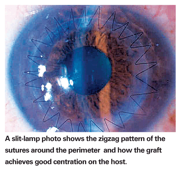
Our approach involves performing PK with a femtosecond laser zigzag incision pattern rather than conventional blade trephination. In our latest published results, the average astigmatism in the zigzag group at three months was 3 D compared with 4.46 D in the group that had the conventional Barron suction trephination PK. Eighty one percent of the zigzag group had best spectacle-corrected visual acuity of 20/40 or better versus 45 percent of the conventional group.1
Problems With Conventional Methods
Under the best circumstances and even in the most skilled hands, full-thickness corneal transplants have been associated with large amounts of regular astigmatism and high-order aberrations. The mechanical blade has a number of inherent issues. Incising the collagen tissue of the cornea naturally induces tissue deformation. The limits of the trephine make it difficult if not impossible to match the incisions of the graft and host cornea.

Surgeons have compensated by oversizing the graft on the host site, but this compounds tissue mismatches and ultimately visual aberrations. The avascular physiology of corneal tissue is fraught with inherent problems for a straight-wall incision. Prolonged healing times, wound separation and leakage, and the need for resuturing are not infrequent complications.
Astigmatism after corneal grafting has two main sources. One is the graft itself. If it does not seat in the same level 360 degrees around, if it is dislocated in one place or pushes forward in another, the warpage and distortion can be significant. Even subtle misalignment undetectable under gross evaluation can be problematic.
The other cause of astigmatism after corneal grafting involves the circumferential sutures. The cornea is more flexible than non-corneal surgeons may realize. Sutures can get bunched on one side and stretch the graft on the other, pulling the graft out into the rim. Thus, meticulous suture placement is paramount to prevent a significant portion of the donor graft settling in the recipient rim. All these factors can translate into optical distortions noticeable to the patient.
Interlocking Graft and Host
Our goal was to improve upon these outcomes by using the more complex and admittedly more costly femtosecond laser. From a patient point of view, the laser offers a few advantages. It can incise tissue without deforming it. It can create a more complex, interlocking pattern between donor and host tissue, which allows more precise tissue apposition than the straight-wall incision and prevents many complications associated with it.
In the laboratory, we used what we called a "top-hat" configuration because a cross section of the incision looked like a top hat. The button had an inner flange with a larger diameter going upward into the corneal stroma. Some surgeons still use this approach and like it, but we found a few problems when we attempted the top hat in human eyes.
The tissue had to be sutured meticulously well because a leak could result if the flange was even slightly out of place. Pushing the flange into place was difficult because we could not exert force on the flexible tissue. The flange did not seal biomechanically. Others have used a "mushroom" approach, basically a reversal of the top-hat pattern.
Advantages of Zigzag Pattern
As we explored other options, we came upon the zigzag pattern. We tried to imitate the inner flap configuration of the clear-cornea cataract incision. We attempted different angles in our early cases and then analyzed the eyes with optical coherence tomography.
This helped us to rapidly modify the parameters. Soon, we achieved the same natural biomechanical properties of a cataract incision in a 360-degree corneal transplant incision.
The zigzag method does not require perfect placement of sutures. Because the sutures do not need to be as tight as other methods, the risk for distortion is reduced. The graft is also inherently self-sealing on the recipient rim because an internal lip creates an interlocking pattern. This provides alignment from front to back. We took the step to make alignment marks on both the donor and recipient tissue as an extra measure of getting perfect alignment.
How Laser Improves Healing
The femtosecond approach, whether using the zigzag, top-hat or a mushroom configuration, provides a broader surface area than conventional mechanical corneal grafting. This translates into a stronger wound, making it safer for the patient in the long run, and allows you to remove the sutures earlier if they induce too much distortion.
We are now evaluating two-year data on patients who have had the zigzag approach, closely examining suture stability. We acknowledge that the cost of using the femtosecond laser for corneal grafting can be an issue for some patients. Medicare does not yet cover this service. Our patients have been enrolled in an internally funded trial, and our goal is to get enough data published to validate the benefit of the laser approach and, ultimately, get a procedure code. Some surgeons code this the same way they do premium intraocular lenses—as an optical service that the patient pays out of pocket—but this can be controversial.
Femtosecond Laser in DALK
We have also described a variation of the big-bubble deep anterior lamellar keratoplasty (DALK) technique using the femtosecond laser zigzag incision.3 As in full-thickness PK, the matching donor and host tissue zigzag cut allows more precise tissue apposition and greater surface area for healing. This also minimizes the risk for perforation of Descemet's membrane. If the big-bubble-Descemet's membrane dissection fails, we can convert to a full-thickness graft while retaining the benefits of the femtosecond laser incision.
Because the DALK technique retains the patient's own endothelium, it eliminates the risk of endothelial graft rejection.
In the meantime, we continue to be encouraged by our results. About half of our patients had VA of 20/25 or better at three months. That is unheard of with previous corneal transplant techniques. We look forward to longer-term results.
Dr. Steinert is professor and chair of the department of ophthalmology, professor of biomedical engineering, and director of the Gavin Herbert Eye Institute,
1. Farid M, Steinert RF, Gaster RN, Chamberlain W, Lin A. Comparison of penetrating keratoplasty performed with a femtosecond laser zigzag incision versus conventional blade trephination. Ophthalmology. 2009 Jul 29 (e pub ahead of print).
2. Farid M, Steinert RF. Results of penetrating keratoplasty performed with a femtosecond laser zigzag incision initial report. Ophthalmology. 2007;114:2208-12.
3. Farid M, Steinert RF. Deep anterior lamellar keratoplasty performed with the femtosecond laser zigzag incision for the treatment of stromal corneal pathology and ectatic disease. J Cataract Refract Surg. 2009;35:809-13.
4. Price FW Jr,


