 |
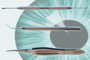
The instruments are the Eippert Femtosecond Spatula, the Solomon Femtosecond Chopper and the LRI Enhancement Forceps. The new devices help surgeons who use femtosecond cataract lasers to create precise subsurface cuts to the eye.
Each offers specific benefits. The Eippert Spatula helps users accurately open primary and secondary incisions created by the femtosecond laser by offering double-ended sizing for greater versatility and blunt, thin tips to maintain proper wound architecture. The Solomon Femtosecond Chopper’s football-shaped tip is the only instrument designed specifically to chop femtosecond-fragmented nuclei. The LRI Enhancement Forceps, which has a 500-µm gauge to correct the depth of incision, can spread accurate incisions during surgery or after, during a slit lamp examination.
For information, call 1 (800) 979-2020, visit
accutome.com or send an e-mail to
info@accutome.com.
New Lotemax 0.5% in a Gel Drop Formulation
Bausch + Lomb announced that Lotemax (loteprednol etabonate ophthalmic gel) 0.5%, which received Food and Drug Administration approval in late September 2012 to treat postoperative inflammation and pain following ocular surgery, will be available January 2013 in pharmacies nationwide. The company says Lotemax is a first-in-class gel drop, with a unique formulation technology. Compared to suspensions, the gel drop formulation is more viscous, allowing it to adhere to the ocular surface.
Another important feature of the Lotemax Gel formulation is that it provides dose uniformity, ensuring that a consistent concentration of loteprednol is delivered in every drop, which is not always possible with corticosteroid suspension formulations. The product is also the only ocular steroid formulation containing glycerin and propylene glycol, two known moisturizers, and has a lower concentration of preservative than Lotemax (lotepredenol etabonate ophthalmic suspension) 0.5% suspension.
In two four-week clinical safety and efficacy evaluations, Lotemax Gel showed statistically significant resolution of anterior chamber cells and flare vs. vehicle at postoperative day eight. Both clinical trials were Phase III, randomized, multicenter, double-masked, parallel-group, vehicle-controlled studies in patients (n=813) being treated for inflammation and pain following cataract surgery. Ocular adverse drug reactions reported in patients treated with Lotemax Gel were eye pain, anterior chamber inflammation, increased lacrimation, photophobia, eye irritation and eye pruritus. Drug-related blurred vision was rarely reported (0.25 percent; 1/407).
For information, visit
bausch.com.
Spectra Iris Indirect Ophthalmoscope from Keeler
Keeler says its new Spectra Iris indirect ophthalmoscope has been specifically designed for portability. Compact and lightweight, the LED indirect has an adjustable aperture for all pupil sizes.
 |
The indirect system is supplied with Keeler’s lightweight wraparound Sport Frames, designed to ensure maximum comfort and balance. It can be worn over glasses and the entire optical unit and light pod can be flipped up to allow direct eye contact when talking to a patient or writing up notes. Spectra Iris can be hung around the user’s neck when not in use, or packed away in its carrying case for storage and transit.
The Spectra Iris can be used continuously for up to four hours on a single battery charge. Its compact lithium ion battery can be clipped onto a belt or stored in its charger when not in use. With a built-in bright, homogeneous LED light source, the need for bulb replacements is eliminated.
Keeler’s Spectra Iris is British-designed and -manufactured. For information, visit
Keelerusa.com, e-mail
Keeler@Keelerusa.com or call 1 (800) 523-5620.
Mobius Therapeutics Announces J Code for Mitosol Mitomycin Solution
Mobius Therapeutics LLC announced today the Centers for Medicare & Medicaid Services has assigned a product-specific Healthcare Common Procedures Coding System (HCPCS) code for Mitosol (mitomycin for solution) 0.2 mg/vial, Kit for Ophthalmic Use. Mitosol is used as an adjunct to ab externo glaucoma surgery. The new J-code, J7315, becomes effective on January 1, 2013.
“This is an important milestone for Mobius Therapeutics, and we are very pleased that CMS has issued a J-code for Mitosol,” said Ed Timm, CEO and founder of Mobius Therapeutics LLC. “While it may take up to three months for the J-code to be loaded into the entire payer system, the J-code facilitates more rapid reimbursement for providers. Mitosol is manufactured under cGMP controls and provides assured dosing concentration with consistent potency and sterility. In light of the on-going concerns related to the sterility and quality assurance of sterile filled medications, Mitosol’s quality manufacturing and convenience helps providers and patients with peace of mind.”
Mobius Therapeutics is a commercial-stage venture focused on ophthalmic surgery solutions. Its first product, Mitosol, is a system for delivering antifibrotic agents in glaucoma, refractive and corneal surgery. The glaucoma indication is in active commercialization; the pterygium and refractive indications are seeking FDA approval. Mobius is housed within the Center for Emerging Technologies in St. Louis, Mo.
For information, visit
mobiustherapeutics.com.
Leica and TrueVision 3D Introduce Digital 3D-Integrated Ophthalmic Microscope
Leica Microsystems and TrueVision 3D Surgical announced that key components of the TrueVision 3D intelligent digital visualization and guidance platform have been integrated with select future models of Leica Microsystems’ ophthalmic surgical microscopes and will be marketed under the Leica brand.
By combining world-class Leica Microsystems optics and illumination with state-of-the-art TrueVision digital stereoscopic imaging, the two companies have partnered to debut a new class of surgical stereo microscope. The companies expect the collaboration to establish integrated 3D visualization and guidance as the standard of care in microsurgery.
The 3D digital integrated microscope can also run TrueVision’s Refractive Cataract Toolset application. The toolset generates precise guidance templates in real-time using preoperative data and advanced algorithms. Surgeons view the 3D live image on the microscope’s 3D HD flat panel display with computer generated overlays for dynamic guidance with eye-tracking during the surgery.
The TrueVision digital 3D system is completely integrated with the Leica M844 and M822 ophthalmic surgical microscopes when equipped with the Leica F40 stand. The system features a patented 10-megapixel HD 3D camera in the optics carrier, 64-bit image processing unit contained within the chassis, and dual passive stereo LED-based LCD displays ranging in size from 23 to 32 inches with articulating arms mounted on the microscope base. The 3D-enabled surgical microscopes are capable of displaying the surgical field of view with 3D guidance and digital overlays on secondary 2D or 3D displays in the operating room.
For information, visit
leica-microsystems.com or
truevisionsys.com. REVIEW


