Sung Bok Lee, MD
C.
Orbital fractures commonly occurby blunt, periocular trauma. Depending on the location and mechanism, intracranial, thoracic and abdominal injuries may be associated. Ophthalmologists most often get involved in pure orbital fractures with an intact orbital rim and without other facial bone fracture. Of orbital fractures, the inferior wall is most commonly involved, followed by the medial wall. The causes of orbital fractures vary, but assault is the most frequent. Other causes include motor vehicle crash, falls (especially in the elderly), sports and industrial accidents.
The Evaluation
A systematically and thoroughly obtained history and physical examination are most important in the evaluation of the traumatized patients. After the identification and treatment of life-threatening injuries, ophthalmologists should rule out serious ocular trauma. Then orbital fractures can be appropriately diagnosed and repaired.
Clinical presentations associated with orbital fractures vary in severity depending on the presence of ocular trauma and the location of the fracture. Symptoms include pain with motility, diplopia with limitation of motion, hypesthesia and trismus. Clinical signs include ecchymosis, crepitus, bone step-off, ptosis, enophthalmos and strabismus. Diplopia and limitation of ocular movement are caused by various conditions. These include orbital hemorrhage and edema, muscular edema or hemorrhage, cranial nerve palsy and entrapment of soft tissue or muscle itself.
Entrapment of tissue occurs in minimally displaced linear or trapdoor fractures, whereas enophthalmos usually occurs in large burst-type fractures. Orbital emphysema is a benign, self-limited condition, but may be aggravated by nose blowing, sneezing or Valsalva maneuver. Oculocardiac reflex may result from entrapment of muscle. It can cause nausea, vomiting and bradycardia, especially in pediatric patients. These symptoms may indicate ischemic damage of the entrapped muscle and suggest immediate surgical intervention.
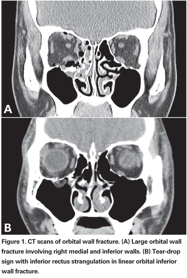
Evaluation of patients with suspected orbital fracture should involve radiologic examination, motility test, diplopia field test and exophthalmometry. Plain X-ray films, although rarely used, with the Caldwell and Waters view may be done as a screening evaluation for possible fractures and foreign bodies. An orbital computed tomography, the gold standard in trauma, CT with contiguous thin axial and coronal sections should be ordered to confirm the diagnosis and plan for treatment (See Figure 1A). Three-dimensional CT reconstruction helps define facial bone anatomy and fractures clearly. Trapdoor fractures in pediatrics may not be noticed in CT scans, and in such cases, tear-drop sign (See Figure 1B), missing rectus sign, severe restriction of motion and oculocardiac reflex can be clues to diagnosing the fractures.

Serial evaluation of the diplopia field test and exophthalmometry are recommended as initial edema, hemorrhage and pain affect evaluation. A forced-duction test may help differentiate the causes of restriction of ocular movement, but one must be careful when interpreting the results. Severe edema or hematoma can mimic entrapment of tissues. The test is useful especially when it is done to judge the entrapment under general anesthesia during surgery.
Conservative observation is recommended when the patients have minimal diplopia with good motility, no evidence of muscle entrapment, no significant enophthalmos, or a small fracture that is unlikely to cause late enophthalmos. Cold compression helps limit orbital edema and hemorrhage. The patient should be told to avoid nose blowing and Valsalva maneuvers, because orbital emphysema and proptosis may result. Oral steroids can be prescribed to speed the resolution of orbital edema and facilitate the surgical decision-making process. Prophylactic antibiotics are suggested if the wound is contaminated, CSF leak is present, or if oral steroids are prescribed.
Surgical repair is recommended if muscle entrapment is suspected, if symptomatic diplopia does not improve over one to two weeks, or if enophthalmos greater than 2 mm is present or anticipated. Because diplopia without muscle entrapment and infraorbital hypesthesia can be resolve with time, these symptoms are not indications for surgery.
When indicated, surgery is generally recommended within two weeks of the injury. If the surgery is delayed, fibrosis between orbital tissues, sinus mucosa and bone fragments makes surgery more difficult. However, some trapdoor fractures may appear well-aligned with minimal edema or hemorrhage, but show marked limitation of motion and oculocardiac reflex. These so-called white-eyed blow-out fractures should be treated immediately to prevent the musculature from ischemic damage and necrosis. If enophthalmos is obvious at the time of presentation and the fracture is large, surgery should not be delayed.
The purpose of surgery is to restore the orbit to its original status before injury. The inferior wall can be easily accessed through transcutaneous or transconjunctival approach (with or without lateral canthotomy). The latter avoids a visible scar and is less likely to result in eyelid retraction. The medial wall can be accessed through transcaruncular approach. Careful exploration under the periosteum allows easy visualization of the fracture boundaries as well as correction of the herniated tissue.
Then various implants can be used to support the orbital soft tissue and prevent recurrent herniation. Porous polyethylene sheets (Medpor) are one of most commonly used implant materials. Other autogenous (cranial, rib or iliac bone graft) or alloplastic (gelatin film, silicone sheet, Teflon, Supramid, titanium mesh or bioresorbable copolymer plates) materials are also available.
Periocular fractures are often managed first by the ophthalmologist. With good clinical examination and radiographic imaging, an informed decision can be made whether surgical intervention is required. For the isolated orbital fractures, the ophthalmologist is well equipped to diagnose and treat these injuries. In the setting of more complex fractures, a multidisciplinary approach may be necessary. However, the ophthalmologist should take the lead as the guardian of ocular function.
Dr. Lee is in the Department of Ophthalmology at
1. Yano H, Nakano M, Anraku K, Suzuki Y, Ishida H, Murakami R, Hirano A. A consecutive case review of orbital blowout fractures and recommendations for comprehensive management. Plast Reconstr Surg 2009;124:602-11.
2. Harris GJ. Orbital blow-out fractures: Surgical timing and technique. Eye 2006;20:1207-12.
3. Burnstine MA. Clinical recommendations for repair of isolated orbital floor fractures. An evidence-based analysis. Ophthalmology 2002;109:1207-13.


