Performing laser refractive surgery on an individual who is in the early stages of keratoconus—or any other corneal thinning disorder—is a bad idea; the consequences of missing this disease can be devastating for both the patient and the surgeon. The problem, as every refractive surgeon knows, is determining whether a patient is headed down that path when the signs of developing disease are still barely detectable.
In this situation, technology is a powerful aid. Every refractive surgeon now scans potential patients using topography and/or tomography, looking for indications that the patient may not be a good candidate. But in many cases, the signs indicating early corneal disease are subtle and ambiguous. Here, three experts share their advice for making the most accurate determination about the presence of a corneal thinning disorder when the evidence produced by this technology is less than conclusive.
Topography or Tomography?
Given the importance of technology in this situation, a key issue is determining which technology is most likely to provide the kind of solid evidence of early disease that surgeons need. Topography, long relied on in this situation, models the anterior corneal surface; tomography provides detailed information about corneal structure.
Stephen D. Klyce, PhD, adjunct professor of ophthalmology at Mt. Sinai University School of Medicine in
At the same time, tomography has proven itself to be extremely valuable. "Probably the biggest thing that's changed recently, in terms of being able to determine whether a patient has forme fruste keratoconus, has been the development of tomography," says Jack T. Holladay, MD, MSEE, FACS, clinical professor of ophthalmology at Baylor College of Medicine in Houston. "Tomography measures slices through the cornea; it's available in instruments such as Oculus's Pentacam, Zeimer's Galilei and Zeiss's Visante Omni, the last being a hybrid of Zeiss's OCT instrument and the Atlas topographer. Tomography not only measures the front surface curvature, but the posterior surface curvature and corneal pachymetry as well. Today, I believe anyone doing refractive surgery needs to have a tomographer for screening."

Dr. Holladay says the changes ac-companying keratoconus are usually more visible on the posterior surface of the cornea. "If you develop an ectasia, the eyelid will move over the bulge about 15 times a minute, 60 minutes an hour, 18 hours a day," he points out.
"Eventually, the epithelium at that location will begin to wear down a little bit. Instead of being six to eight layers thick, it ends up being only four or five layers thick. The result is that the elevation on the front of the cornea is not as great as the elevation on the back.
That's why the changes you find on the posterior surface are much more sensitive indicators of change than the changes on the anterior surface."
According to Dr. Holladay, the Pentacam, Galilei and Visante Omni all pick up posterior surface changes quite well. One way they do so is by comparing the posterior corneal surface to a hypothetical toric ellipsoid as a reference surface. "If the posterior surface is more than 10 µm elevated above that toric ellipsoid reference surface, that's keratoconus," he explains. "If it's less than 5 µm elevated above the reference surface, that's normal. If it's between 5 and 10 µm, it's in the gray area where you can't be sure. These devices are all set up to make this kind of comparison."
Apparently, most surgeons today are not relying on topography alone. "At a recent ISRS symposium in
The Power of Pachymetry
Of course, when screening for corneal thinning disorders, measuring corneal thickness is important. "In addition to surface changes, corneal thickness changes from the norm can also indicate the presence of disease," observes Dr. Klyce. "Most data suggest that a cornea less than 500 µm is abnormally thin. The traditional way to evaluate corneal thickness is with an acoustic pachometer, by taking central, superior, inferior, temporal and nasal readings. Those five readings are usually adequate for finding areas in which the cornea is thinner than usual."
Of course, many of the more recent instruments can also measure corneal thickness. "Tomographers can measure the posterior surface of the cornea, and combining that information with elevation data from the anterior surface can provide pretty good estimates of corneal thickness across the cornea, from limbus to limbus," says Dr. Klyce.
"That's very handy as a way to look for thin spots, especially inferiorly with keratoconus. You can also use a global pachymetry map to look for the radial distribution of corneal thickness. Some published data suggest that there's a correlation between keratoconus and how thickness changes from the center to the edge."1
In terms of technology, devices using Scheimpflug imaging may be advantageous. "From a clinical point of view, these devices give a good overview of the tomographic information, and they're quick and easy enough to be used for screening," Dr. Belin says. "An instrument that gives you every possible bit of information but requires a special technician and takes 30 minutes to perform a scan is not very useful."
Dr. Klyce warns, however, that the technology isn't flawless. "In my experience, the Orbscan is the only device that, by itself, is fairly reliable for both topography and thickness measurements," he says. "The Pentacam doesn't have a placido disc for measuring topography; the Galilei has the placido disc and the Scheimpflug scanning device, but it merges the placido data and elevation slit data, which can degrade the sensitivity of the placido data.
"A few surgeons are using only the Pentacam for screening corneas, but I believe that's a mistake," he continues. "The Pentacam displays a corneal topography map, but the measurements are actually generated from tomographic data, based on corneal shape and elevation. Because of the way the topographic data is derived, a placido device can be as much as 20 times more sensitive to topographic contours than a tomographer."
Despite the greater topographic accuracy of placido technology, Dr. Klyce notes that in some situations tomography is more advantageous for measuring the topographic component. "Tomographic devices like the Pentacam are very good at analyzing corneas that are very distorted or aberrated," he notes. "That includes corneas with keratoconus or ectasia. That's because on very distorted corneas, placido devices have lots of data dropout. The rings, or mires, tend to get very close together and finally merge when a cornea is steep or distorted. Ideally, a clinician would use a placido device to get very sensitive anterior surface measurements, and a tomographer, such as a scanning Scheimpflug device, for analyzing more distorted corneas."
Choosing the Right Color Scale
When interpreting a scan accurately is crucial, the way the data is represented onscreen can make a huge difference. Dr. Klyce and his team developed the color-coded contour map that's now the international standard for displaying corneal topography.2 "There are about 10 topographers on the market, and every one of them tries to be different from the others," he says. "They all have a selection of scales and ways to represent corneal power. It becomes very confounding for the clinician. In the end, many clinicians pick the default scale or play around with the options, trying to make sense of it.
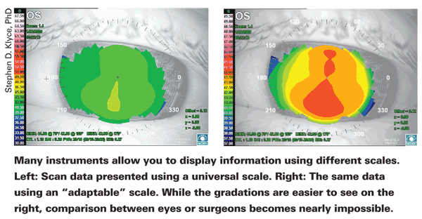
"Unfortunately," he continues, "it's very difficult to determine what's normal and abnormal if you don't use a fixed, standard scale; and it's almost impossible for one clinician to compare information with another. Furthermore, many topographers are shipped with a so-called 'adaptable' scale, in which every cornea is presented with its own color scale. This complicates matters further." (See sample maps, above.) He notes that the international standard scale he helped develop is available in most topographers today. "That's my number one pearl," he says. "Use the fixed, standard scale."
Dr. Belin agrees. "Adaptive or relative scaling is probably not the way to go," he says. "If you change scales constantly you can't make comparisons over time, and you can't make a quick visual inspection because maps may look identical while the information they contain is totally different."
The Relative Pachymetry Map
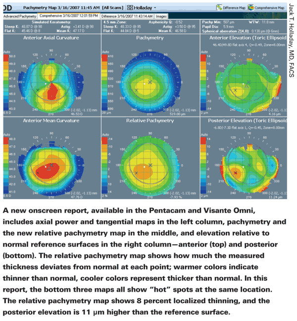
As any weather buff knows, there are many ways to integrate and present available information, and the way you choose to analyze and present it can make all the difference in how useful it is at the practical level. In that spirit, Dr. Holladay says he's helped to develop a new map that displays key information in a novel way, called the relative pachymetry map.
"Standard pachymetry maps are normally red in the center; the colors get cooler as you move toward the periphery because the cornea gets thicker," he notes. "With this kind of scale, if you have a little thinning in the area where the cone is, it may be difficult to see. For example, let's say a cornea is 555 µm in the middle and 700 µm in the periphery, and the thickness is 630 µm in the area of the cone, where it should be 650 µm. This is still thicker than 555 µm, so with this sliding color scale the cone won't show up very clearly on the map as an area of suspicion.
"The new relative pachymetry map uses a different approach," he continues. "We know the normal thickness of the cornea and how much thicker it becomes as you move away from the center. So, we changed the scale to reflect what percent of that normal thickness is found at each point. A completely normal map would be all green. If the measured thickness is 10 percent thinner than normal at a given point, a warmer color at that point indicates -10 percent. If it's 10 percent thicker, a cooler color indicates +10 percent. So the map shows whether—and by how much—the corneal thickness deviates from normal. Any deviation greater than 3 percent is significant, and anything over 6 percent is absolutely too thin."
Taking this idea further, Dr. Holladay has helped to develop new onscreen reports for the Pentacam and the Visante Omni that incorporate this information. "The screen shows six maps, arranged in three columns," he says. "In the first column the report shows the axial power map at the top and the tangential map at the bottom. The middle column shows the pachymetry map on top and the relative pachymetry map on the bottom. On the right, the elevation of the front surface relative to the reference surface is on top, and the posterior surface relative to its reference surface is at the bottom.
"All the clinician has to do is look at the bottom row," he continues. "If you see a hotspot on the tangential map, that indicates inferior steepening.
Then you look at the relative pachymetry map and say, aha, that same spot is a little thinner than it should be. Then you look at the posterior float map that compares the elevation to the toric ellipsoid. If it's greater than 10 µm in that area, that's three strikes —you're out. The patient has keratoconus and shouldn't have LASIK."
Pearls for Practice
The following strategies may help you make the tough calls in day-to-day practice:
• Always do corneal topography first. "Putting in dilating drops, touching the cornea to measure IOP or manipulating the eye for a long period of time will cause disruption or modification of the corneal tear film," explains Dr. Klyce. "That's a problem, because corneal topography measures the shape of the air-tear interface. That's where the greatest refractive index change occurs, and that's the surface from which the placido disc is reflected. Many times you'll see a topography scan that looks a little irregular, and you'll notice that the pupil is 8 mm wide. In that situation it's pretty clear that the technicians started the dilating drops before they did corneal topography."
• For routine clinical screening, use the axial diopter corneal power display. "There are two other ways to present corneal power: refractive power or instantaneous tangential power," says Dr. Klyce. "Both of those are useful for other purposes, but the axial diopter corneal power display is best for routine screening."
• Look for changes in the posterior surface that are greater than the changes in the anterior surface. "I've never seen a case of keratoconus in which the changes on the posterior surface did not exceed—and also predate—the changes on the anterior surface," notes Dr. Belin. "In my experience, the posterior surface and the pachymetric distribution are early indicators of pathology."
• With a younger patient, check recent spectacle prescriptions. "The person that's the most difficult—and most important—to catch is the 21-year-old patient who comes in for laser surgery with early keratoconus," says Dr. Holladay. "That person's condition is the hardest to detect because he's so young that the keratoconus hasn't really had a chance to manifest yet. However, on tomography you may note some against-the-rule astigmatism and inferior steepening that's very mild, slight changes in the relative pachymetry map and posterior elevation map—suspicious, but not diagnostic. What should you do?
"It's a good idea to ask for the patient's spectacle prescriptions for the past four or five years," he says. "Almost all patients with keratoconus end up with changes in their refraction, especially when they're young. If the patient has early keratoconus, you'll see changes in the astigmatism correction in his glasses that reveal increasing asymmetrical against-the-rule astigmatism. In contrast, most people who have high degrees of astigmatism but no keratoconus have close to the same amount of astigmatism in each eye over time, and it's symmetrical."
• Look for asymmetry in the scan. "Keratoconus is usually defined as a localized area of steepening on corneal topography," notes Dr. Klyce. "This can occur anywhere on the corneal surface, and it has several faces. It can look like an asymmetric bow tie, for example. (A symmetric bow tie is normal, regular corneal astigmatism.) Keratoconus also can show up as a truncated bow tie—a very short, steep bow tie in the center of the cornea. It can also resemble a moderate amount of corneal astigmatism, but with skewing of the radial axis, so the bow tie looks a little bit weepy or saggy. (See sample map, p. 24.)
"Not every corneal topographer contains a good screening program to differentiate normals from abnormals, so clinicians should at least learn how to estimate what's called the I-S value," he adds. "This is an index that's used to look for abnormally high amounts of asymmetry. If it's elevated, look for other signs of keratoconus."
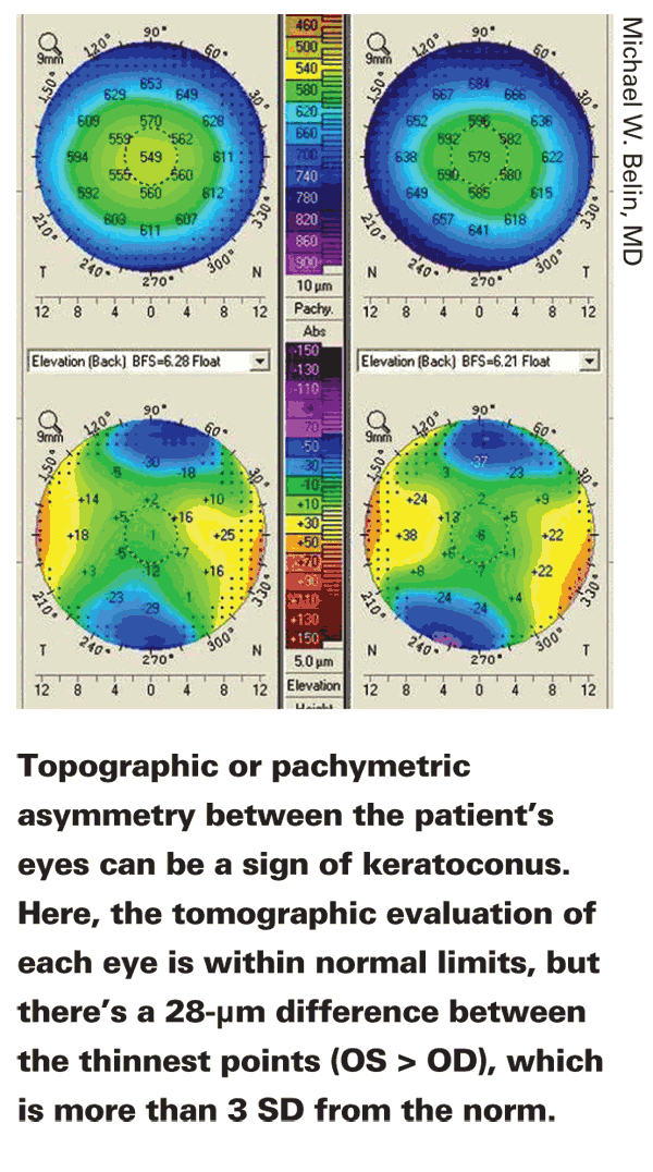
• Check for topographic or pachymetric asymmetry between the two eyes. "This is another sign of keratoconus, as well as some other conditions," says Dr. Klyce. "If one eye has with-the-rule astigmatism and the other eye has against-the-rule astigmatism, you need to look for other signs of keratoconus. Also, check for differences in central corneal thickness between the two eyes. The two eyes should be symmetrical in both corneal topography and pachymetry. If they're not, that's generally a sign of abnormality and a risk factor for developing complications after refractive surgery." 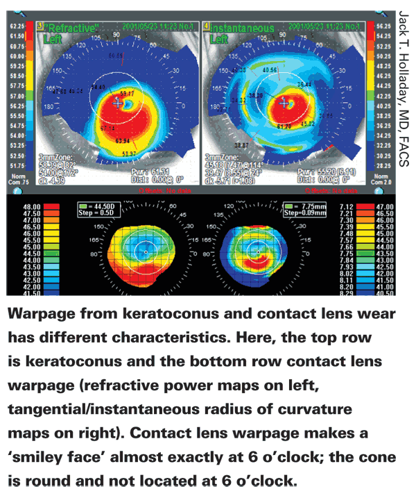
Dr. Belin agrees. "Dr. Stephen S. Khachikian and I published a study two years ago evaluating pachymetric asymmetry between fellow eyes," he says.3 "In medicine, we know that even when numbers are normal, asymmetry often indicates potential disease. For example, if someone has an intraocular pressure of 9 mmHg in both eyes, we'd think little of it. If the pressure is 18 in both eyes, we probably wouldn't think twice about it, as long as everything else was normal. But if the pressure is 9 in one eye and 18 in the other, that's a red flag.
"Our study found that a 23.2-µm difference in pachymetry at the thinnest point of the two eyes represented three standard deviations from the norm," he continues. "That means that highly asymmetric maps should raise a cautionary flag."
• If a topographic scan looks odd, check the visible cornea image with the mires reflecting. "When the computer processes placido concentric ring reflections, you may sometimes find errors because of low contrast, steep areas or other problems," notes Dr. Klyce. "Most topographers have a utility that allows you to look at the image of the cornea with the mires present; it lets you see if there's mistracking of the rings, and whether that mistracking is in the area of abnormal steepening or abnormal flattening. If that's the case, you can discard that exam, or at least be aware that the abnormality is an artifact and not use it in your evaluation."
• Take the patient's age into account. "A normal cornea cross-links by itself as we age, gradually becoming more rigid," notes Dr. Holladay. "When we're young, the cornea changes shape easily. By the time we get to age 60 or 70, it's more like plastic. So if a 20-year-old shows signs of keratoconus, his risk is much greater than that of a 50-year-old with the same signs."
• Don't just look at the information you're accustomed to getting from earlier technology. "Sometimes clinicians have machines that are capable of giving them a lot of information, but they just look at the information they could get from their old machines," says Dr. Belin. "Make sure you read all the instructional information that comes with your device."
• If you encounter a map that you can't decipher, get help. Dr. Holladay says he gets three to 10 e-mails a day from doctors asking for help interpreting scans. "If you don't know what's going on," he says, "ask for help from someone who has more experience and learn from him."
• Know how to check your in-strument's calibration. "Although today's machines are sturdy, it is possible for them to be knocked out of calibration," observes Dr. Klyce.
"One clinic had a week in which every patient appeared to have inferior steepening. It turned out that the cleaning crew had bumped the cone on the topographer and dislodged it a little one night.
"If you're routinely using an instrument, scan your own eye periodically," he says. "Your techs should become familiar with the topography of their own eyes or that of a colleague. If it changes markedly, then something is wrong and the unit needs to be looked at. Or, if scans all begin to seem suspicious, you can use the calibration surface that comes with the instrument."
Managing Contact Lens Patients
Because wearing contact lenses is well-known as a source of corneal distortion, having the patient stop wearing the lenses before making final measurements for surgery is crucial. How long should the surgeon wait? "Everyone has their own guidelines regarding how long to keep that patient out of lenses," says Dr. Belin. "That decision also somewhat depends whether it's a soft contact lens, toric lens or rigid lens. Many surgeons, if it's a spherical soft lens, would wait only a few days. But if the lenses are rigid, the patient will need to be out longer. If a map shows distortion or abnormalities, keep the pa-tient out of the lenses and repeat the map until you get a stable reading."
"With most modern soft and rigid gas permeable contact lenses, it takes two to four weeks for the effect of the lenses on the cornea to dissipate," says Dr. Klyce. "We usually recommend that patients discontinue wear for a couple of weeks before screening. If the corneal shape is abnormal—for example, if we find inferior steepening that mimics keratoconus but is actually contact lens warpage—we ask patients to keep their lenses out and come back in two to three weeks, until the topography normalizes."
Because contact lenses and early disease can both cause corneal changes, differentiating between them is important. These strategies may help:
• Determine on which surface the changes are occurring. "It's well-known that contact lens wear can induce changes on the anterior surface," says Dr. Belin. "We've also seen changes on the posterior surface in some cases. How can we tell if warpage is caused by the contact lens? The only sure way is to discontinue lens wear for a while and see what the cornea does. However, I've never seen a case of keratoconus in which the posterior surface changes didn't exceed those on the anterior surface. With contact lenses, you commonly see the changes on the anterior surface. Finding the latter is one indicator that this may be contact lens warpage."
• Compare the eyes. "If the warpage is contact lens induced, the distortion in both eyes will be symmetrical," says Dr. Holladay. "If it's keratoconus, they won't look like mirror images—one will be worse than the other."
• Note the characteristics of the inferior steepening. "The changes in inferior steepening caused by contact lens wear are always exactly at 6 o'clock, and they're always symmetrical," Dr. Holladay points out. "In keratoconus, changes in inferior steepening are never at 6 o'clock and never symmetrical. (See sample maps, facing page.) If the patient has keratoconus and stops wearing his contacts for a few weeks, you'll see the cornea begin to show the position and asymmetry signs of keratoconus."
• Compare the thickness of the cornea and cone. "If a cone is contact lens-induced, the cornea will be thicker at that point due to epithelial hyperplasia," notes Dr. Holladay. "If it's keratoconus, the cornea will be thinner there than it should be. So, take an ultrasound probe and measure over the cone, and then at a symmetrically opposite point above the pupil. If the patient has keratoconus, the cornea will be thinner by more than 20 µm. If not, the cornea will be the same, or thicker below."
Reasons for Hope (and Caution)
For a refractive surgeon, identifying early keratoconus is obviously crucial for protecting both the patient and the surgeon from serious postop trouble. But today, there are other reasons to uncover keratoconus in its early stages as well: Improvements in treatment may make it possible to save these patients from what used to be inevitable consequences of the disease.
"John Kanellopoulos in Greece is taking people with keratoconus, doing topographic-guided wavefront to get rid of the bump and improve their optics, and then doing corneal cross-linking," notes Dr. Holladay. "He now has 10 to 12 years of follow-up, and these people are actually getting better. So not only is our ability to diagnose the disease improving, the treatment is improving to the point that we can, in essence, cure the disease." He notes that cross-linking isn't available in the
Despite this extra reason to identify individuals who may be developing keratoconus, the chief concern on the minds of most refractive surgeons is still avoiding a postoperative disaster. And the reality is that deciding whether to proceed with surgery can be difficult.
"I've seen cases in which the corneal topography really did look normal, with no ectasia, but the postop result was corneal irregularities," says Dr. Klyce. "So complications can happen, even when you practice the most conservative, highest level, best-trained style of medical care. My advice is to always to be conservative with refractive surgery.
RO
"At the same time," he adds, "there's a lot of economic pressure today; no one wants to turn away an eager patient who appears to be healthy. It can be a tough decision."
Dr. Klyce is a consultant for Nidek. Dr. Holladay is a consultant to Zeiss and Oculus. Dr. Belin is a consultant to Oculus, but has no financial stake in the company.
1. Ambrósio R Jr, Alonso RS, Luz A, Coca Velarde LG. Corneal-thickness spatial profile and corneal-volume distribution: Tomographic indices to detect keratoconus. J Cataract Refract Surg 2006;32:11:1851-9.
2. Smolek MK,
3. Khachikian SS, Belin MW, Ciolino JB. Intrasubject corneal thickness asymmetry. J Refract Surg 2008;24:6:606-9.