Determining the exact etiology and best treatment for blepharitis often seems to raise more questions than answers. In fact, even though the disease is very common, we're not much closer to cracking its mysteries than we were 40 years ago, so don't expect this article to be filled with life-altering revelations. However, if you'd like to be brought up to date on what we actually know about blepharitis and the best ways to classify and treat it, read on.
Etiology
The apparent incidence of blepharitis in the general population is high, and blepharitis diagnoses in the ophthalmologist's office are frequent. Even though it's common, however, our understanding of the disease's underlying pathophysiology is incomplete. Also surprising is the lack of clear-cut definitions as to what constitutes the global category of blepharitis. Because of these shortcomings, blepharitis therapy remains somewhat primitive, and isn't directed at the underlying etiology.
 |
| Occlusion of the meibomian glands with a keratinized sheet can occur in blepharitis. |
Historically, the rule with blepharitis cases was that a third were caused by Staphylococcus, another third by seborrhea, and the final third were from a combination of Staph and seborrhea, mixed with causes such as Demodex folliculorum mites, Pityrosporum ovale, or fungi. Angular blepharitis, a distinct entity, is often caused by Staphylococcus and moraxella, or is associated with allergic neurodermatitis.
The use of certain drugs can cause blepharitis. Some patients on cancer chemotherapeutic agents such as 5-fluorouracil have been documented as having ocular surface and lacrimal complications, including blepharitis, conjunctivitis, keratitis and eyelid dermatitis.1 The association of systemic retinoids, such as isotretinoin (Accutane) used for acne vulgaris treatment, has been documented to result in tear-film and ocular-surface problems arising from the drying property of the medication. Clinical studies have found blepharitis in 40 to 55 percent of patients following isotretinoin treatment. In addition, one study showed that the percentage of patients culturing positive for S. aureus in the conjunctival sac jumped from 7.3 percent before treatment to 61.8 percent during it. Tear-film breakup time decreased in 69.1 percent of patients in the same study.2,3
The reason for these changes with isotretinoin administration is the drug's ability to decrease meibomian gland function, subsequently affecting the osmolarity of tears and allowing for excessive evaporation of the tear film.4 This relationship between isotretinoin and the development of blepharitis warrants further study, as it may offer insights into the mechanism and etiology of blepharitis.
Blepharitis's association with seborrheic dermatitis can't be neglected.5 Interestingly, blepharitis is often seen in conjunction with rosacea (more likely ocular, but also dermatologic), which is itself a disease of unknown etiology.6
Researchers have also noted that there is a relatively high incidence of blepharitis in patients with Down's syndrome. One study evaluating 152 children with Down's syndrome found 30 percent had blepharitis. In addition, 30 percent had a lacrimal system obstruction.7
It's clear that many etiologies of blepharitis exist, and therefore, as we'll discuss later, it's certain that one form of therapy would not be sufficient for all of them.
A Review of Signs and Symptoms
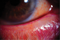
• Signs. The hallmark sign of blepharitis is lid margin redness. Upon slit-lamp exam, this redness is typically accompanied by an apparent increase in the subepithelial vascular network, as well as a change from the sharp, angled lid margin to a rounder, more curved margin. The presence of collarettes around the base of each lash is also a key sign. If one pulls on a collarette, it becomes clear that it's rooted within the lid itself, giving evidence for a layer of translucent hyperkeratinized tissue.
Alterations in the meibomian glands include changes in their contents. Their clear oil will turn opaque, progressing to a thick paste, or a gland itself can become covered by an operculum. The alterations eventually result in scarification and a shifting and loss of the linear arrangement of the glands along the lid margins. Lid notching and subepithelial fibrosis may also exist. One study found that 74 percent of chronic blepharitis patients had meibomian gland dropout compared to 20 percent of normals.8
Tear-film abnormalities are a prominent feature in meibomian-gland dysfunction (MGD). In contact lens wearers, MGD is also often present and is important to watch for, not only with regard to blepharitis, but because it can make lens wear intolerable. In patients with MGD, one study has shown higher rates of tear evaporation.9 Further study has noted normalization of tear breakup time if the deep contents of the meibomian glands are added to the tear film.10 As mentioned, the etiology may include rosacea or seborrheic dermatitis, so noting these, and their frequency of association with chalazia, can be an important diagnostic tip-off.
• Symptoms. The symptoms of blepharitis commonly include irritation and a tickling/itching sensation. It's important to note that the itching of blepharitis is different from that of allergy. While there is an allergic blepharitis caused by allergens or drug-induced allergy that can often involve the lid margin, the itching associated with true blepharitis is distinct from that seen with seasonal allergic conjunctivitis. The itching with blepharitis is more of an intermittent tickle, rather than the severe itch of allergy that makes patients feel the need to vigorously rub or scratch.
Categorization
Given our poor understanding of blepharitis, and since its etiologies are so varied, definition and categorization of the disease and its forms are challenging. Several different categorizations for blepharitis have been proposed, based on various criteria. Here's a look at them.
The categorization of blepharitis devised by James McCulley, MD, of the University of Texas Southwestern Medical Center represents the best clinical approach to date. It separates the disease into six groups:11
• Staphylococcal. This is marked by acute inflammation of the lids. This group consists mainly of women, many also having dry eye.
• Seborrheic. This is characterized by the involvement of clusters of meibomian glands and the presence of scales with an oily appearance near the lashes.
• Combined staphylococcal and seborrheic involvement.
• Seborrheic with secondary meibomian seborrhea. In this, the meibomian secretions are too profuse.
• Seborrheic blepharitis with secondary meibomian gland inflammation. This results in thickening of meibomian secretions and blocking of the gland ducts.
• Primary meibomitis with dermal involvement (i.e., acne rosacea or seborrheic dermatitis). This is marked by an unstable tear film and severe signs and symptoms.
Another classification scheme relies more on meibomian gland involvement (i.e., seborrheic with meibomian-gland dysfunction, obstructive meibomian-gland dysfunction, obstructive with dry eye, and dry eye alone).
A third scheme uses anatomical distinctions, taking a broader look at blepharitis and characterizing it based on two simple principles: the presence of staphylococcal infection in anterior lid margin blepharitis and the presence of meibomian-gland dysfunction in posterior lid margin blepharitis.12 While its simple distinction is helpful, this method doesn't offer the specific distinctions of the six-group system.
It's important to recognize that the meibomian glands themselves may be affected in ways distinct from changes in vascularity or loss of lid-margin contour. In the lid-margin metaplasia of dry eye and rosacea, cause and effect are uncertain. Instead, the metaplasia is like that seen with ectropion, in which the conjunctiva keratinizes once it's no longer within the tear film.
Treatment
Devising the proper treatment plan for blepharitis can be challenging, and multiple possible etiologies contribute greatly to the difficulty. This is also a condition that relies heavily on the patient as a partner in achieving disease management, since lid scrubs and hot compresses can require high levels of long-term compliance to produce positive results.
Underlying conditions should be treated first. Dry eye is highly likely, and it can only help to take the first step by getting this under control. In addition, underlying seborrhea should be treated (See sidebar, "Seborrheic Dermatitis," below). In fact, treating the scalp may even help treat the lids.
The use of warm compresses and lid scrubs can be crucial in controlling blepharitis. These aren't cures, though. Warm compresses placed on the lids several times a day, followed by massaging of the lids, can help break up blockage of meibomian gland ducts and stimulate secretions. Lid scrubs help reduce lid-margin debris and eliminate bacteria, and can be done after warm compresses.
In cases resulting from staphylococcal infection, antibiotics are typically effective, including systemic antibiotics such as tetracycline (250 mg q.i.d.) or doxycycline (50 mg b.i.d.). Not only do these antibiotics result in significant decreases in bacteria on the eyelids, it appears they also have positive lipid-enhancing effects on the tear film. Experts believe that these effects are induced by a lipase-inhibiting property of these drugs.13-15 Be mindful of adverse reactions with antibiotics, however, as both tetracycline and doxycycline can cause gastrointestinal complaints and sun sensitivity. Avoid using them in children, or women who are pregnant or nursing.
The use of a topical ophthalmic steroid or antibiotic can be helpful in reducing acute inflammation. With steroids, it's important to be aware of the potential for steroid-related complications and to use them for the shortest duration possible. Antibiotic-resistant bacteria now pose a threat to the treatment of blepharitis. However, the latest generation of ophthalmic antibiotics, the fourth-generation fluoroquinolones, has been able to maintain bactericidal efficacy.
Much about blepharitis remains a mystery. Simply knowing the complexity of the systems involved makes it amazing that its prevalence isn't greater.
However, looking ahead, there's hope from such researchers as Dr. McCulley and his colleagues, who continue to conduct exemplary studies.
We're acutely aware of the prevalence of blepharitis in our practices, and knowing that we don't know all of the answers will drive us all to learn all we can about this disease.
Dr. Abelson, an associate clinical professor of ophthalmology at Harvard Medical School and senior clinical scientist at Schepens Eye Research Institute, consults in ophthalmic pharmaceuticals.
Mr. Cohane and Ms. Fink are research associates at Ophthalmic Research Associates in North Andover.
1. Eiseman AS, Flanagan JC, Brooks AB, Mitchell EP, Pemberton CH. Ocular surface, ocular adnexal, and lacrimal complications associated with the use of systemic 5-fluorouracil. Ophthal Plast Reconstr Surg 2003;19:3:216-24.
2. Bozkurt B, Irkec MT, Atakan N, Orhan M, Geyik PO. Lacrimal function and ocular complications in patients treated with systemic isotretinoin. Eur J Ophthalmol 2002;12:3:173-6.
3. Egger SF, Huber-Spitzy V, Bohler K, Raff M, Scholda C, Barisani T, Vecsei VP. Ocular side effects associated with 13-cis-retinoic acid therapy for acne vulgaris: clinical features, alterations of tear film and conjunctival flora. Acta Ophthalmol Scand 1995;73:4:355-7.
4. Mathers WD, Shields WJ, Sachdev MS, Petroll WM, Jester JV. Meibomian gland morphology and tear osmolarity: Changes with Accutane therapy. Cornea 1991;10:4:286-90.
5. Gupta AK, Bluhm R, Cooper EA, Summerbell RC, Batra R. Seborrheic dermatitis. Dermatol Clin 2003;21:3:401-12.
6. Ghanem VC, Mehra N, Wong S, Mannis MJ. The prevalence of ocular signs in acne rosacea: Comparing patients from ophthalmic and dermatology clinics. Cornea 2003;22:3:230-3.
7. Da Cunha RP, Moreira JB. Ocular findings in Down's syndrome. Am J Ophthalmol 1996;122:2:236-44.
8. Mathers WD, Shields WJ, Sachdev MS, Petroll MW, Jester JV. Meibomian gland dysfunction. Cornea 1991;10:4:277-285.
9. Mathers WD. Ocular evaporation in meibomian gland dysfunction and dry eye. Ophthalmology 1993;100:3:347-51.
10. McCulley JP, Sciallis GF. Meibomian keratoconjunctivitis. Am J Ophthalmol 1977;84:6:788-93.
11. McCulley JP, Dougherty JM, Deneau DG. Classification of blepharitis. Ophthalmology 1982;89:1173.
12. Smith RE, Flowers CW. Chronic blepharitis: A review. CLAO Journal 1995;21:3:200-207.
13. Ta CN, Shine WE, McCulley JP, Pandya A, Trattler W, Norbury JW. Effects of minocycline on the ocular flora of patients with acne rosacea or seborrheic blepharitis. Cornea 2003;22:6:545-8.
14. Shine WE, McCulley JP, Pandya AG. Minocycline effect on meibomian gland lipids in meibomianitis patients. Exp Eye Res 2003;76:4:417.
15. Dougherty JM, McCulley JP, Silvany RE, Meyer DR. The role of tetracycline in chronic blepharitis: Inhibition of lipase production in Staphylococci. Invest Ophthalmol Vis Sci 1991;32:2970-5.


