A 50 year-old Caucasian female with sudden onset of blurred vision, floaters and metamorphopsia in her right eye presented for ophthalmic evaluation. Four to five days prior to onset of the visual symptoms, she developed headache, fatigue and flank pain, followed by patches of hives on her torso and lower extremities. She presented to a local emergency room where a CT scan showed mesenteric adenitis. She denied any symptoms in her left eye. Of note, the patient had recently suffered a deep scratch from a stray cat she had adopted.
Medical History
Past ocular history included LASIK in both eyes. Past medical history disclosed arthritis, chronic kidney disease and sinusitis. Family history included a father who had heart disease. Social history was significant for owning a stray cat but was otherwise unremarkable.
Current medications included: artificial tears; conjugated estrogens/bazedoxifene; levocetirizine; aspirin; vitamin D; vitamin E; and fish oil.
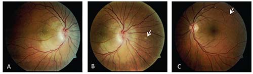 |
| Figure 1. Fundus photos of the right eye’s macula (A) and optic nerve (B) demonstrating optic disc edema with extensive peripapillary edema, macular edema, and hard exudates. The left eye (C) is normal with the exception of a white retinal lesion, also seen in the right eye (arrows). |
Examination
Ocular examination demonstrated visual acuity of count fingers in the right eye and 20/50-2 in the left eye. Pupils were pharmacologically dilated at the time of examination. Intraocular pressures were 9 and 10 mmHg in the right and left eyes, respectively. Confrontation visual fields were diffusely constricted in both eyes. Extraocular motility was full bilaterally. The anterior segment examination revealed intact LASIK flaps and trace nuclear sclerosis of the lens bilaterally but was otherwise unremarkable.
Dilated fundus examination of the right eye demonstrated trace vitreous cell, hyperemic optic disc edema with extensive peripapillary edema, macular edema with hard exudates and venous tortuosity (See Figure 1). The left eye had a normal optic disc, macula and vessels. Small white lesions were observed in both eyes, nasal to the optic disc in the right eye and at the superior vascular arcade in the left eye (Figure 1, arrows).
Click here for diagnosis, workup and discussion.


