Quite possibly no other complication fills LASIK surgeons with as much dread as corneal ectasia. The desire to avoid it has led to the development of high-tech topographic indices designed to catch keratoconus suspects, intricate preop risk-assessment algorithms and hours of lectures and research presentations at every major ophthalmology meeting. Having a patient develop ectasia was akin to dropping off a cliff, an inevitable fall that ended in corneal transplantation. Now, however, researchers may have developed a parachute: corneal cross-linking. Cross-linking, which aims to chemically strengthen corneal tissue to arrest, or in some cases reverse, the progress of ectasia is showing promise in studies in the Unites States and abroad. Here's a look at the results surgeons are achieving with cross-linking and its associated treatments.
A Link to a Cure?
The proposed mechanism of corneal cross-linking is a strengthening of the structural stability of the cornea by fostering cross-links between, or in some cases inside, collagen fibers using a combination of riboflavin and ultraviolet light.1 This strengthening may freeze ectasia and keratoconus progression.
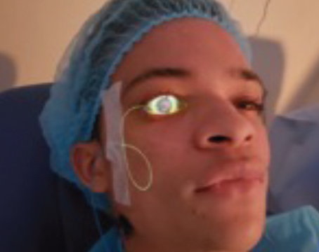
Though the cross-linking technique may vary slightly from one study to the next, it usually proceeds as follows: Under topical anesthesia, the surgeon debrides the epithelium of the ectatic cornea out to 7 or 8 mm. He then applies riboflavin for a period of five to 15 minutes before the irradiation step, giving the agent time to penetrate the cornea and eventually appear in the anterior chamber. At that point, the physician begins to irradiate the cornea with ultraviolet-A light at a surface irradiance of 3 mW/cm2 for a period of 30 minutes. During this time, he continues to apply riboflavin every two to three minutes. Afterward, the physician instills a topical antibiotic and places a bandage contact lens to protect the cornea until it can reepithelialize. Some physicians will also send the patient home with additional topical medications. "There's a moderate amount of pain after the treatment," says
Preop, we give patients a systemic non-steroidal anti-inflammatory such as 600 mg ibuprofen and a couple of drops of tetracaine and lidocaine jelly. Postop, we send them home with a systemic NSAID, a narcotic analgesic for use for the first day or so to avoid any significant pain and the topical antibiotic. We also give them some topical NSAIDs, such as Acular LS, primarily for the first week to help control discomfort, as NSAIDs can have some topical anesthetic effect."
The FDA Trial
The only randomized, controlled FDA clinical trial analyzing the safety and efficacy of cross-linking for post-surgical ectasia is being sponsored by the German company Peschke-Meditrade, and is currently recruiting and treating patients at 10 centers. Peschke-Meditrade is the distributor of the UV-X, a UV emitter that's used for cross-linking. The study is also designed to look at the treatment's effect in keratoconus.
In the ectasia arm of the study, the trial will include 160 patients with ectasia who will be randomized to the treatment or control group. In the treatment group, one eye receives the cross-linking. After three months, patients in the control group are allowed to cross over into the treatment group.
R. Doyle Stulting, MD, PhD, the study's director, says the three-month results have been positive, and seem similar to the results experienced by various European cross-linking studies. "The treatment patients are doing better than the controls, with a slight decrease in corneal curvature at three months. But it's important to note that the goal of the treatment is to stop progression, not cause regression. So, any regression that we get is an added benefit." Dr. Stulting says both ectasia patients and keratoconus patients respond to the treatment in a similar way. The average K values at one, three and six months appear in Figure 1. In 45 patients at three months, the average K reading had decreased by 0.29 D, while it increased slightly in the control group.
In the study, visual acuity is a secondary outcome measurement. "Right now, there is no difference in that outcome measure, but it might be too early with too few patients for us to see that," explains Dr. Stulting.
The treatment has resulted in a couple of side effects so far. "A number of the patients get a little bit of haze, probably as a result of the treatment and cross-linking, but [this is] not a true adverse event," he says. "We've had a couple of cases of infectious and non-infectious corneal ulcers, as you might expect from a treatment where there's an epithelial defect and then contact lens wear as the defect heals. They healed without any visually significant scarring." In the entire study, there was one culture-positive and one culture-negative infiltrate, and two that weren't cultured. Four eyes had delayed re-epithelialization and one developed uveitis. Dr. Stulting is planning to present data on a greater number of patients at this year's meeting of the
Some might wonder if one cross-linking treatment will be enough for these patients. However, European data, as Dr. Stulting points out, appears to show that the effect persists. In a trial from
Athens
Prophylactic Cross-linking
While it would be a great help to have a treatment for postop ectasia, some surgeons think it might be even better to prevent ectasia from ever occurring by performing cross-linking as an adjunct to refractive laser surgery in high-risk patients. Dr. Kanellopoulos has performed prophylactic cross-linking on 150 LASIK and PRK cases over the past three years, and says the eyes have remained stable so far.
He uses the technique on patients that require a high LASIK correction, which he defines as over -8 D with relatively thin corneas (500 to 520 µm) or any LASIK cornea with an irregularity that's not definitely keratoconus. "The LASIK is performed routinely, with at least a 300-µm residual stromal bed," Dr. Kanellopoulos explains. "Then, prior to putting the flap back in place, I instill several drops of riboflavin 0.1 phosphate sodium on the stromal bed and the underside of the flap. One minute later, and after achieving clear visualization of the riboflavin in the stroma and the flap, I reposition the flap, irrigate it and fit it into place with a
Dr. Kanellopoulos studied his prophylactic technique in 25 high-risk cases and, at an average follow-up of 1.5 years, the average K-readings went from 44.5 to 36.25 D, uncorrected acuity improved from 0.2 (Snellen: 20/100) to 1.00 (Snellen: 20/20), best-corrected acuity went from 0.9 (Snellen: a little better than 20/25) to 1.2 (Snellen: just under 20/16), with no cases of ectasia.
Linking Up with Intacs
Another approach some surgeons have taken when faced with ectasia is the use of intracorneal ring segments, both as a stand-alone treatment and in combination with cross-linking.
Efekan Coskunseven
The implantation of the corneal rings is performed under sterile conditions and topical anesthesia. Dr. Coskunseven uses the Purkinje reflex as the central point and marks it under the WaveLight Allegretto Biomicroscope. He uses a 5-mm marker to locate the exact ring channel and measures the corneal thickness intraoperatively using ultrasonic pachymetry along the ring location marks. He sets the tunnel depth at 80 percent of the thinnest corneal thickness at the tunnel location.
Dr. Coskunseven bases the arc length and thickness of the segments on the manufacturer's nomogram, and he uses a 60-kHz femtosecond laser to create the channels. The channels' inner diameter is set to 4.4 mm, and the outer diameter is 5.6 mm.
The entry cut thickness is 1 µm, and the ring energy used for channel creation is 1.30 µj.
He implants the intracorneal ring segments immediately after channel creation, before the bubbles disappear, as Dr. Coskunseven says these bubbles show the exact tunnel locations. "To avoid any injury to the incision area, the segments are directly implanted with the special KeraRing forceps," he says.
Postoperatively, patients instill antibiotic and steroid drops q.i.d. for two weeks. Dr. Coskunseven instructs them to avoid rubbing their eyes and to use preservative-free artificial tears frequently. On the first postop day, he performs a slit-lamp exam to evaluate corneal wound healing and to look for any migration of the segments.
"Incomplete tunnel creation is one the most common complications of femtosecond laser channel creation because tissue bridges in the tunnel may cause problems during ICRS implantation," explains Dr. Coskunseven. "My advice is to increase the energy level of the femtosecond laser or decrease the spot separation in order to avoid such complications."
If the ICRS implantation is to be combined with cross-linking, Dr. Coskunseven follows the post-ICRS implantation results out to six months. "If the K readings are greater than
1 D, we perform cross-linking to stop the progression and achieve further improvements in visual and refractive outcomes," he says. If the patient's younger than 20, has a corneal thickness less than 400 µm with the risk of superficial migration of the segments or can't return for follow-up, he'll perform cross-linking on the worse eye one month after segment implantation, and then perform cross-linking on the fellow eye a month after that.
In a small study of his technique, Dr. Coskunseven took 16 ectatic eyes of 10 patients and performed ICRS implantation on 10 eyes, ICRS/cross-linking on four and cross-linking alone on two. At an average follow-up of nine months, the combination patients' uncorrected vision had improved from a preop value of 0.14 (Snellen: a little worse than 20/125) to 0.35 (Snellen: a little worse than 20/50). Best-corrected vision improved from a preop of 0.27 (Snellen: slightly better than 20/80) to 0.6 (Snellen: near 20/32). The average K reading improved from 52.4 D to 48.45 D.
Cross-linking appears to be a boon for surgeons, and future randomized studies will help define the procedure's indications. "It's a treatment for diseases that have no other treatment," says Dr. Stulting. "And, if applied early, it has the potential to prevent progression of either keratoconus and ectasia to the point that contact lenses or corneal transplants are required."
1. Hafezi F, Kanellopoulos J, Wiltfang R, Seiler T. Corneal collagen crosslinking with riboflavin and ultraviolet A to treat induced keratectasia after laser in situ keratomileusis. J Cataract Refract Surg 2007;33:2035-2040.
2. Wittig-Silva C, Whiting M, Lamoureux E, et al. A randomized controlled trial of corneal collagen cross-linking in progressive keratoconus: Preliminary results. J Refract Surg 2008;24:S720-S725.