The natural history and economic impact of this disease is devastating, as most patients with untreated exudative AMD will become legally blind within two years of diagnosis. In addition to the loss of financial resources and reduced quality of life, it is estimated that advanced AMD is responsible for a 0.3-percent reduction of the United States gross domestic product, or $30 billion.
Monotherapy
Prior to the availability of anti-vascular endothelial growth factor agents in early 2005, the only available treatment options for choroidal neovascularization secondary to AMD were destructive interventions such as thermal laser and photodynamic therapy with verteporfin. Treatment with thermal laser, as guided by the Macular Photocoagulation Study, remains an option for some extrafoveal and juxtafoveal lesions. However, the high risk of recurrence and permanent blind spot may make this an unappealing option in the modern era.4-6
A number of clinical trials testing PDT demonstrated a slowing of visual loss in some patients with CNV. Patients in the Treatment of Age-Related Macular Degeneration with Photodynamic Therapy (TAP) study had classic CNV, and showed a benefit in visual stabilization.7,8 A small subset of that cohort, patients with pure classic CNV, did significantly better than those with predominantly classic disease, and statistically drove the efficacy for the entire group.
Two early trials, VIP (Visudyne In Photodynamic Therapy) and VIM (Visudyne in Minimally Classic), suggested that PDT might be beneficial in patients with occult CNV.9,10 Based on these trials and the lack of other available treatment options, the Centers for Medicare & Medicaid Services (CMS) extended reimbursement of PDT for occult CNV in many cases, although it remains unapproved by the Food and Drug Administration for eyes with occult CNV. The Visudyne in Occult (VIO) trial did not achieve the primary end point, suggesting that PDT is not effective in occult CNV.11 Many retina specialists have developed significant experience with PDT, and based on the combination of trial results and clinical experience, continue to use PDT, especially in combination with other therapies.
The FDA approved pegaptanib (Macugen, OSI/Eyetech), a pegylated aptamer that blocks VEGF 165, in December 2004 for the treatment of all subtypes and locations of exudative AMD. This broad approval was based on the VEGF Inhibition Study in Ocular Neovascularization (VISION) trials, which demonstrated a significant benefit in reduction of visual loss and visual gain in patients with all forms of exudative AMD.12 In the VISION trial, 70 percent of eyes injected with Macugen every six weeks for one year achieved visual stabilization, compared to 55 percent of controls. This statistically significant finding ushered in the anti-VEGF therapy era, and more than 50,000 patients have been injected with Macugen. Both in the trial and in clinical practice, multiple injections of Macugen seem to be extremely well-tolerated, and the selective blockade of VEGF-165 may provide a safety advantage over more broad-spectrum anti-VEGF agents. Subsequent analyses suggest that treating eyes with early onset of CNV enhances the efficacy of Macugen.13
Bevacizumab (Avastin, Genentech) is a full-length anti-VEGF antibody that is FDA-approved for intravenous use in the treatment of colon cancer. Retina specialists have utilized it extensively over the past year to treat both AMD and retinal vascular diseases. While a number of papers have been written describing utilization of the drug, no prospective clinical trials have been completed. While the drug clinically appears to be effective and safe, the true efficacy and safety of Avastin in AMD remains unknown. Although the use of Avastin for AMD is off-label, many Medicare carriers reimburse its use in AMD.
On June 30, 2006 the FDA approved ranibizumab (Lucentis, Genentech), an anti-VEGF antibody fragment, for the treatment of all forms of exudative AMD. In two pivotal phase three trials, MARINA (Minimally classic/occult trial of the Anti-VEGF antibody Ranibizumab In the treatment of Neovascular AMD) and ANCHOR (ANti-VEGF Antibody for the Treatment of Predominantly Classic CHORoidal Neovascularization in AMD), approximately 95 percent of patients maintained or improved their vision. These eyes were treated monthly with intravitreal Lucentis, and the FDA label reflects that treatment regimen. Lucentis is the major breakthrough in AMD management to date, as it is the first treatment that restores vision, reflected in the 35 percent of eyes that gained three or more lines in these trials. In addition, nearly 40 percent of eyes treated in the trials achieved a visual acuity of 20/40 or better.
Testing
The accurate evaluation and assessment of CNV continues to be a critical component of successful AMD management. Ophthalmoscopic examination and fundus drawing remain important to the evaluation process, as both non-contact and contact methods are vital when trying to synthesize the results of imaging tests. Fluorescein angiography (FA) provides important data concerning wet-AMD. While the days of arguing over whether CNV is classic or occult seem somewhat antiquated, the differentiation is still helpful prognostically. FA remains vital in identifying lesions with retinal angiomatous proliferation, pigment epithelial detachment, and feeder vessels. In addition, FA is indispensable in assessing CNV growth.
Optical Coherence Tomography is utilized both for diagnosis of AMD and monitoring response to treatment. OCT can demonstrate actual CNV complexes, including elevation, thickening and fibrosis of the retinal pigment epithelium. In addition, cystoid macular edema (CME), subretinal fluid and retinal thickening can be identified and quantified. Pre-treatment anatomic status is important to document, as anatomic changes from baseline can be utilized to monitor therapeutic effect.
Many physicians choose to use OCT primarily when evaluating therapeutic success with AMD therapy. FA is just as critical for monitoring patients after treatment. While OCT is an excellent imaging modality, reproducibility of images, fixation in eyes with central field loss and inability to gauge lateral growth of CNV remain major challenges. In multiple trials such as ANCHOR and MARINA, OCT anatomy has not corresponded to visual acuity. Because of these issues, we continue to utilize FA extensively in AMD management. Indocyanine green angiography, both standard and high-speed, can provide information concerning location and amount of leakage, and identify lesion type and extent.
Early detection and intervention in AMD is critical to maximizing outcomes. Education of the general public, and other eye care professionals, is a critical component to successful management of AMD.
Integration
Integration of the multiple treatment options for AMD illustrates the age-old adage that medicine is both an art and a science. While diagnosis of exudative AMD is oftentimes not complicated, decisions concerning choice of therapeutic agent, treatment interval, frequency and cessation, combination therapy and cost of intervention often are. These complicated issues contribute to significant variations in practice patterns between equally competent physicians. Strong opinions about therapeutic regimens and excellent public debates have made this an exciting time in retinal practice.
Our clinical approach centers around the utilization of evidence from clinical trials to guide therapeutic decisions. We attempt to integrate testing results with treatment options to guide our practice. On initial evaluation, we perform ophthalmoscopy, OCT and FA. It is important to identify parameters for each individual patient that can be followed to evaluate the efficacy of treatment. Visual acuity, angiographic leakage, OCT anatomy and clinical appearance are parameters that can be monitored. Conflicting findings on these evaluations can be confusing, but nonetheless are important. Each case may show variation in response, and adequate time needs to be allowed for a given treatment to modify a given parameter.
In cases of small peripapillary membranes and extrafoveal CNV, argon laser may be offered, especially in patients with financial restrictions or difficulty traveling for frequent visits. Peripapillary membranes may also be observed, especially if not near the fovea. We often utilize one injection of Avastin or Lucentis two weeks before the laser therapy in order to shrink the CNV and minimize the area that needs to be treated. Reduced subretinal fluid also allows for adequate burns to be achieved with lower laser power. This may or may not influence the rate of recurrence and scar extension. We are currently evaluating the use of anti-VEGF agents after laser to maintain CNV inactivity.
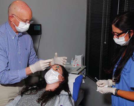
Unfortunately, most patients have a subfoveal component of their CNV at time of presentation. These patients are ideally started on either Lucentis or Avastin. Significant debate exists concerning the benefits of both drugs. Those arguments are beyond the scope of this review. That being said, we typically prefer Lucentis, as it is FDA-approved, safe and has excellent efficacy. Avastin is also a very good option, but because of the lack of prospective trial data, most of our patients choose to utilize Lucentis.
In the ANCHOR and MARINA trials, patients gained most of their vision during the first four injections of drug. Therefore, we encourage our patients to commit initially to four injections, given at one-month intervals. At the end of that period, we re-evaluate with OCT and FA. At this point, we review alternative treatment strategies including data from the PIER (A Phase IIIb, multicenter, randomized, double-masked, sham injection-controlled study of the efficacy and safety of ranibizumab in subjects with subfoveal choroidal neovascularization with or without classic CNV secondary to AMD) and PrONTO (Prospective Optical coherence tomography imaging of patients with Neovascular age-related macular degeneration Treated with intra-Ocular Lucentis) trials. In light of the reduced efficacy of the PIER protocol, which evaluated induction with three monthly injections of Lucentis followed thereafter by doses once every three months, we discourage a strategy of planned injections.
The PrONTO study is evaluating an alternative treatment strategy, where patients are given three monthly injections of Lucentis, and are retreated if the patient demonstrates an increase in central OCT thickness of at least 100 µm, a loss of five letters in conjunction with recurrent fluid by OCT, a new onset of classic neovascularization, or a new macular hemorrhage. A PrONTO-style protocol is certainly attractive to both patients and clinicians. However, we find ourselves having to temper our enthusiasm for the preliminary results of PrONTO, as the study is a small, open-label, uncontrolled single investigator trial. In light of this, we strongly encourage monocular patients to follow an evidence-based approach and continue with monthly injections, while binocular patients are asked to participate in the decision-making process more vigorously.
We usually do not repeat ancillary testing during the first three months unless patients are worsening clinically. In eyes with classic CNV, PDT is considered if the patient does not respond to Lucentis after three doses. PDT is not typically given in eyes with occult CNV in light of the VIO results. Other considerations include the addition of intravitreal Kenalog, which may act in synergy with anti-VEGF agents.
It is challenging to define a true failure of either Lucentis or Avastin. If a patient fails one of these drugs, changing them to the other medication is reasonable in light of the significant differences in half-life, size, and affinity for VEGF. The problem is that outcomes data for the trials are at a one-year time point. Interim data on OCT and FA is not available. Many patients on Lucentis or Avastin will show a visual response during the first three to four injections, but may not show an anatomic response, and continue to have OCT and FA evidence (particularly the latter) of CNV activity. This is a theme seen with other anti-VEGF medications, such as pegaptanib. One may ask whether a patient who appears active clinically but has a visual improvement or stability has failed the therapy. This is a difficult issue to manage, and we typically take an approach based on the best evidence available.
Treated patients typically fall into one of three clinical scenarios. The first group is eyes that have had a visual response but not an anatomic response. Patients in this group are kept on monthly Lucentis injections. The second group is patients who have anatomic response but not visual response. These patients are presumably suffering from photoreceptor damage or fibrosis that occurred during the period of CNV activity. Patients in this group are offered a PrONTO style management strategy.
The third group is patients who have neither anatomic nor visual response after four injections. These patients can reasonably have their drug changed or be started on combination therapy. Nonetheless, we do feel that these patients should continue to receive some anti-VEGF agent, unless significant fibrosis has developed. It is not clear if patients who initially do poorly might recover at one year with continued therapy. As a result, we usually err on the side of retreatment with monthly injections to give patients the best chance of achieving the MARINA and ANCHOR results. Adding PDT may or may not be beneficial, and based on results of the FOCUS trial (RhuFab V2 Ocular Treatment Combining the Use of Visudyne to Evaluate Safety), it is not. Cross trial comparison is always difficult, but a lower percentage of patients in FOCUS achieved visual stabilization when compared to those in MARINA and ANCHOR.
With the intense media coverage of these new drugs, management of patient expectations has become challenging. Many patients enter the office expecting a miracle cure for their disease, even if they have years of advanced vision loss. While patient input on therapeutic strategy is important, the burden of what strategy to employ is placed on the physician, and the patient needs to use physician-directed advice to help make that decision. We all must do our best to educate patients so that they understand the benefits and limitations of the current treatment options. Although anti-VEGF agents are effective, they do not cure all cases of wet-AMD. Abnormal neovascular vessels do not completely resorb with anti-VEGF therapy, and other growth factors continue to stimulate activity. Many lesions faced in clinical practice are more advanced than those enrolled in trials, and these more mature CNV may not be responsive to current therapies.
The future management of AMD will involve even more therapeutic options. Bevasiranib (small interfering RNA), VEGF trap, small molecules and implantation of stem cells under the retina, may add to our armamentarium. While these new treatments will ultimately bring about further benefits to our patients, they will also add even more choices in the management of this complex disease.
Dr. Halperin is a partner in the Retina Group of Florida, in Boca Raton. Dr. Prenner is an assistant clinical professor at the Retina Vitreous Center of the Robert Wood Johnson Medical School, UMDNJ, in New Brunswick, NJ.
1. Friedman DS, O'Colmain BJ, Munoz B, et al. Prevalence of age-related macular degeneration in the United States. Arch Ophthalmol 2004;122:564-572.
2. Klein R, Klein BE, Linton KL. Prevalence of age-related maculopathy. The Beaver Dam Eye Study. Ophthalmology 1992;99:933-943.
3. Clemons TE, Milton RC, Klein R et al. AREDS Study Group: Risk factors for the incidence of Advanced Age-Related Macular Degeneration in the Age-Related Eye Disease Study (AREDS). AREDS report no. 19. Ophthalmology 2005;112:533-9.
4. Macular Photocoagulation Study Group. Argon laser photocoagulation for neovascular maculopathy; five-year results from randomized clinical trials. Arch Ophthalmol 1991;109:1109-1114.
5. Macular Photocoagulation Study Group. Laser photocoagulation of subfoveal neovascular lesions in age related macular degeneration; results of a randomized clinical trial. Arch Ophthalmol 1991;109:1220-1231.
6. Macular Photocoagulation Study Group. Laser photocoagulation for juxtafoveal choroidal neovascularization; five-year results form randomized clinical trials. Arch Ophthalmol. 1994;112:500-509.
7. Treatment of Age-Related Macular Degeneration With Photodynamic Therapy (TAP) Study Group. PDT therapy of subfoveal choroidal neovascularization in age-related macular degeneration with verteporfin; one year results of 2 randomized clinical trials, TAP rport 1. Arch Ophthalmol 1999;117:1329-1345.
8. Bressler NM. Photodynamic therapy of subfoveal choroidal neovascularization in age-related macular degeneration with verteporfin; two-year results of 2 randomized clinical trials –TAP report 2. Arch Ophthalmol 2001;119:198-207.
9. Verteporfin in Photodynamic Therapy Study Group. Verteporfin therapy of subfoveal choroidal neovascularization secondary to age-related macular degeneration; two year results of a randomized clinical trial including lesions with occult with no classic choroidal neovascularization-VIP report 2. Am J Ophthalmol 2001;131:541-560.
10. Gragoudis ES, Adamis AP, Cunningham ET Jr. et al for the VEGF Inhibition Study in Ocular Neovascularization Clinical Trial Group. Pegaptanib for neovascular age-related macular degeneration. N Engl J Med 2004;351:2806-2816.
11. Gonzales CR, Adamis AP, Cunningham ET Jr, et al. Enhanced efficacy associated with early treatment of neovascular age-related macular degeneration with pegaptanib sodium: An exploratory analysis. Retina 2005;25:815-827.