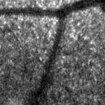
The prototype instrument, simply called the polarimetric scanning laser ophthalmoscope, collects images by emitting fixed, linear polarized laser light into the eye at a wavelength of 785 nm. On the output side, there's an analyzer that consists of devices called a linear polarizer and a quarter-wave plate. This setup enables the system to produce images of retinal blood vessels with improved contrast than that possible without the polarization techniques. The researchers have imaged three eyes so far, but haven't yet compared their device to commercially available ones.
"There are commercial instruments that use light's polarization properties," says Brian Vohnsen, PhD, one of the researchers behind the polarimetric SLO. "They usually make images of 20 degrees of the retina, whereas we have scanned very small areas down to about 2 degrees. These commercial instruments have two orthogonal polarizers in their setup, so we're looking at different combinations of polarization." He says the system can more or less double the contrast in images of retinal blood vessels compared to images without these polarization techniques.
"The main application of the system would be glaucoma diagnosis," explains Dr. Vohnsen. "If you can get a higher resolution of a small area, you can see changes at an earlier stage than you could with present diagnostic systems. You could look at small areas of the rim of the optic disc, for example." At this point, though, he admits, "But since we're not monitoring what part of the changes come from the cornea or the lens, it's hard to say if you're seeing a retinal change."
He says the system's current resolution is stopping them from going in the direction of differentiating the polarization from the various ocular structures, and therein lies his future development task with the SLO. "We have the problem that our resolution still isn't good enough, so we're thinking about working adaptive optics into the system," he says. "We'd like to get a resolution down to a few microns, so you could have essentially a diffraction-limited imaging of the retina." By diffraction limited, he means that the limiting factor of the resolution is the effect of diffraction of light, not any imperfections in the device's optics.
"We'd also like to look at more eyes where we can find some more interesting features and not just look at blood-vessel contrast."
 |
 |
| Two images from the polarimetric SLO. The left image is the original image with the best polarization detection, and the other has been processed to improve the blood vessel contrast. |


