Presentation
A 58-year-old man noted progressive vision loss in both eyes. He was seen by a retina specialist and found to have subretinal fluid in the right eye suggestive of central serous chorioretinopathy. He was initially followed for one month but developed progressive vision loss in the right eye with subsequent involvement of the left. His symptoms didn’t improve with one injection of intravitreal bevacizumab, and he was referred to the Ocular Oncology Service at Wills Eye Hospital for a second opinion.
Medical History
Six months prior to presentation, he was diagnosed with stage IV esophageal cancer with nodal metastasis involving the supraclavicular, mediastinal and retroperitoneal region. He had completed five cycles of docetaxel for the malignancy. Additional medications included dexamethasone, prochlorperazine, lorazepam, oxycodone and granisetron. Past ocular history was notable for a retinal detachment in the right eye that had been repaired with surgery 12 years prior. Family history was noncontributory and social history was notable for being a former smoker. He was allergic to ciprofloxacin.
Examination
On ophthalmic examination the uncorrected visual acuity was counting fingers with no pinhole improvement
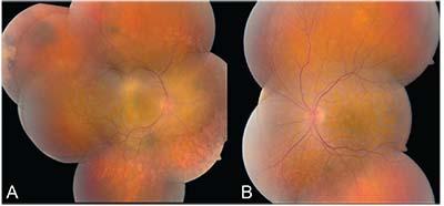 |
| Figure 1. Montage color fundus photograph of the right eye (A) and left eye (B) revealing bilateral multifocal subtle pigmented choroidal lesions and diffuse nummular retinal pigment epithelial loss, submacular and peripheral geographic orange lipofuscin pigment, and subretinal fluid. |
in the right eye and 20/40 with no pinhole improvement in the left. The pupils were equal, round and reactive to light without afferent pupillary defect. Applanation tonometry revealed intraocular pressure of 17 mmHg in the right eye and 12 mmHg in the left eye.
External examination was unremarkable. Anterior segment examination revealed symmetric conjunctival injection and bilateral cataract with moderate nuclear sclerosis, and anterior and posterior subcapsular opacification in both eyes.
On dilated fundoscopic examination, both eyes had multifocal subtle pigmented choroidal lesions. There were six lesions in the right eye, ranging from 1 mm to 6 mm in diameter, and there was extensive subretinal orange pigment, particularly nasally and inferonasally (Figure 1A). The left eye showed similar but less advanced findings with three pigmented choroidal lesions, all measuring 1 mm or less in diameter with diffuse overlying orange pigment (Figure 1B).
Click here for Diagnosis & Discussion.


