Relevant Anatomy
The confluence of the eyelid skin and tarsal conjunctiva forms the eyelid margin. At this mucocutaneous junction lies the “gray line,” formed by a specialized strip of pretarsal orbicularis, first described by the French anatomist Jean Riolan in the 17th century.1 The eyelashes, numbering approximately 100 in the upper lid and 50 in the lower, originate anterior to the tarsus, pass between the gray line and the remainder of pretarsal orbicularis, exiting the skin at the anterior lid margin. The meibomian gland orifices are aligned between the lashes and the gray line.
Etiopathogenesis
Any condition causing scarring of the tarsal plate and the conjunctiva can cause lash misdirection. The causes can be categorized as follows:
1) Trauma
• Eyelid injury
|
• Thermal burns to the face, lids
• Post-surgical changes, e.g., ectropion repair
2) Infections
• Trachoma. Trachoma is the leading infectious cause of blindness worldwide and overall is the eighth-commonest blinding disease.2 It is currently endemic in 57 countries, most of which are in Sub-Saharan Africa and Asia.3 Trachomatous trichiasis is the result of multiple infections with Chlamydia trachomatis since childhood, resulting in chronic inflammation and scarring of the tarsal conjunctiva, pathognomonically called the Arlt’s line.
• Herpes zoster. Eyelids are commonly involved in herpes zoster ophthalmicus. Patients may develop blepharitis, leading to secondary bacterial infections, scarring and trichiasis.
3) Autoimmune diseases
• Ocular cicatricial pemphigoid. This is a subclass of mucous membrane pemphigoid affecting predominantly the conjunctiva. OCP is a Type II hypersensitivity response with antibodies binding at the basement membrane zone, leading to the activation of complement and recruitment of inflammatory cells. The release of cytokines causes fibroblast activation with consequent progression to scarring.
• Stevens-Johnson syndrome. This is a severe cell-mediated hypersensitivity reaction to drugs or antigens modified by drug exposure. SJS causes severe ocular morbidity. The acute phase is characterised by severe hemorrhagic lid crusting, pseudomembranous or membranous conjunctivitis, which progress to severe cicatrisation of the lids and conjunctiva in the late stages.
4) Inflammation
• Blepharitis. Long-standing cases of blepharitis result in scarring of the eyelid.
• Vernal Keratoconjunctivitis. This is a recurrent bilateral IgE-mediated disorder, primarily involving the upper tarsal conjunctiva.
|
Clinical Picture
Symptoms: Patients may be asymptomatic or may have a range of symptoms which include foreign body sensation, redness, lacrimation, pain and photophobia.
Signs: A thorough slit-lamp examination of the anterior segment is to be performed in every patient to assess the distribution of trichiatic eyelashes, elucidate the underlying cause and rule out differential diagnoses. Table 1 lists diagnostic clues that will aid in getting to the root cause of the trichiasis.
Although trachoma is uncommon in the United States, the general ophthalmologist might come across a large number of cases of trachoma during service trips abroad or in recent immigrants from endemic areas. It is the responsibility of each and every ophthalmologist to identify and treat every case of trachomatous trichiasis, to ensure that the WHO goal to eradicate blinding trachoma by 2020 becomes a reality.
Differential Diagnosis
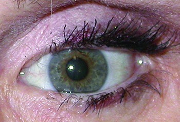
1) Pseudotrichiasis. As mentioned earlier, pseudotrichiasis occurs in the setting of lid malposition, including entropion (See Figure 2) and epiblepharon.
• Entropion. Involutional changes are the most common cause of entropion and primarily affect the lower eyelid. Rarely, entropion can occur due to conditions that cause cicatrisation and shortening of the posterior lamella like trachoma, OCP and other bullous diseases, chemical injuries, radiotherapy and trauma. Functional entropion can be tested by checking for lid laxity and asking the patient to close the eyes forcefully, noting rotation of lashes towards the globe.
|
• Epiblepharon. This is a congenital lid anomaly in which the skin and the pretarsal orbicularis override the lid margin, causing the lashes to be directed vertically against the ocular surface. It is common in children of Asian origin and noticed shortly after birth.
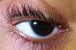
2) Distichiasis. Distichiasis is a rare disorder defined as the abnormal growth of lashes from the orifices of the meibomian glands on the posterior lamella of the tarsal plate causing an aberrant second row of lashes (See Figure 3). It can be congenital or acquired. The congenital form occurs when a primary epithelial germ cell destined to differentiate into a meibomian gland of the tarsus develops into a pilosebaceous unit. It can be isolated or associated with ptosis, strabismus, congenital heart defect or mandibulofacial dysostosis. In the acquired form, there is metaplasia and dedifferentiation of the meibomian glands into hair follicles, secondary to conditions like severe chemical burn, SJS, OCP or chronic blepharoconjunctivitis.
Management of Trichiasis
No diagnostic procedures are required in the management of trichiasis. However, conjunctival biopsy can be performed if trachoma or SJS is suspected.
Medical management: Treatment of trichiasis is primarily surgical. Medical measures are aimed at controlling the symptoms and treating the underlying condition.
• Lubricants, such as artificial tears and ointments, provide relief from the irritant effects of lash rubbing.
• Immunomodulatory therapy and supportive systemic therapy are used in OCP and SJS.
• In cases of trachoma, antibiotics should be administered to those affected and to all family members. A single dose of azithromycin (20 mg/kg up to 1 g) is the treatment of choice.
• A single dose of azithromycin also reduces postoperative trichiasis recurrence rates by one third compared to topical tetracycline in trachoma-endemic areas.4
• Doxycycline may prevent the recurrence of trichiasis following surgery in patients with trachoma by altering matrix remodelling and contraction by conjunctival fibroblasts.5
Surgical Management
Many procedures have been described for the treatment of trichiasis.
• Mechanical epilation with forceps is a simple temporary method of removing misdirected lashes, but the lashes grow back in three to six weeks. Broken cilia are often more irritating to the cornea than mature long eyelashes. Despite these drawbacks, epilation is cheap and is generally acceptable to patients as a treatment modality.6
• Electrolysis using a high frequency electrical current is a useful procedure for few isolated lashes, but has a number of drawbacks. It has a high recurrence rate, causes scarring of the adjacent eyelid margin and can be tedious for the patient as well as the surgeon.
• Radiofrequency ablation of lashes and follicles is a simple yet effective method which can be performed under local anaesthesia. A small gauge wire is introduced alongside the lash down to the follicle. The radiofrequency signal is delivered for about 1 second with the lowest power setting on cut/coag mode to destroy the hair follicle. Application of 0.02% mitomycin C in conjunction with radiofrequency ablation may help to improve the success rate of radiofrequency ablation treatment.7
|
• Laser ablation.
A) Argon laser photocoagulation can be performed to ablate a few isolated lashes. In this, the laser is fired into the depths of the eyelid, at the base of the errant lash to cauterise the follicle, which is then pulled out. Hence, this procedure works best in pigmented individuals. Complications include mild hypopigmentation and lid notching.8
B) The 810 nm diode laser is an effective tool in the treatment of trichiasis.9 The pulse length used is around 50 ms and the energy intensity is approximately 50 J/cm2. The patient might require four to five sittings four to six weeks apart.
C) Epilation with a ruby laser is also a viable and well-tolerated option for the relief of symptoms of trichiasis.10
• Trephination is a new technique in which the lash follicles are cut with a 1.0 mm microtrephine.11 This is a safe, effective, fast technique with fewer complications and less scarring.11
• Surgery involving full-thickness pentagonal resection or anterior lamellar rotation excision may be performed for segmental trichiasis resistant to other methods of treatment.
• A simple surgical procedure in which the anterior lamella is removed, and the eyelid is allowed to heal by spontaneous granulation, has been described for severe trichiasis.12
Management of Pseudotrichiasis
• Entropion. Strengthening of the lower lid retractors (Jones procedure) and lateral tarsal strip can be used for cases with horizontal lid laxity. Tarsal fracture with repositioning of the anterior lid margin and mucous membrane grafts are techniques for posterior lamellar scarring causing cicatricial entropion.
• Epiblepharon. Spontaneous resolution with age is the rule. Hotz procedure (excising a strip of skin and muscle below the lid margin and fixing the skin crease to the tarsal plate) can be performed for persistent cases.
Management of Distichiasis
Mechanical epilation, electrolysis, radiosurgery or cryosurgery can be used to take care of the aberrant second row of lashes. However, unlike trichiatic lashes, in distichiasis the cilium shaft runs a circuitous course from the bulb to the surface, making destruction with an epilating needle unpredictable. Internal lash resection beneath a skin muscle flap or combination of lid-splitting and cryotherapy may be effective. REVIEW
Dr. Kleinberg is the Orbit and Skull Base fellow at Wills Eye Hospital. She will be joining Worcester Ophthalmology Associates in Worcester, Mass., this summer. Dr. Srinivasan is an ophthalmologist from India, and is currently a research fellow at Wills Eye Hospital.
1. Mackie IA. Riolan’s muscle: Action and indications for botulinum toxin injection. Eye 2000;14:347-352.
2. Resnikoff S, Pascolini D, Mariotti S, Pokharel G. Global magnitude of visual impairment caused by uncorrected refractive errors in 2004. Bull World Health Organ 2008;86:63-70.
3. Mariotti S, Pascolini D, Rose-Nussbaumer J. Trachoma: Global magnitude of a preventable cause of blindness. Br J Ophthalmol 2009;93:563-568.
4. West S, West E, Alemayehu W, Melese M, et al. Single-dose azithromycin prevents trichiasis recurrence following surgery: Randomized trial in Ethiopia. Arch Ophthalmol 2006:124:309-314.
5. Li H, Ezra D, Burton M, Bailly M. Doxycycline prevents matrix remodelling and contraction by trichiasis-derived conjunctival fibroblasts. Invest Ohthalmol Vis Sci 2013;54:4675-4682.
6. Rajak S, Habtamu E, Weiss H, Kello A, et al. Surgery versus epilation for the treatment of minor trichiasis in Ethiopia: A randomised controlled noninferiority trial. PLoS medicine 2011;8:e1001136.
7. Kim G, Yoo W, Kim S, Han Y, et al. The Effect of 0.02% Mitomycin C Injection into the Hair Follicle with Radiofrequency Ablation in Trichiasis Patients. Korean J Ophthalmol 2014;28:12-18.
8. Al-Bdour M, Al-Till M. Argon Laser: A Modality of Treatment for Trichiasis. Int J Biomed Sci 2007,3:56.
9. Pham R, Biesman B, Silkiss R. Treatment of trichiasis using an 810-nm diode laser: an efficacy study. Ophthal Plast Reconstr Surg 2006; 22:445-447.
10. Moore J, De Silva S, O’Hare K, Humphry R. Ruby laser for the treatment of trichiasis. Lasers MedSci 2009;24:137-139.
11. McCracken M, Kikkawa D, Vasani S. Treatment of trichiasis and distichiasis by eyelash trephination. Ophthalmic Plastic & Reconstructive Surgery 2006, 22(5), 349-351.
12. Moosavi A, Mollan S, Berry-Brincat A, Abbott J., et al. Simple surgery for severe trichiasis. Ophthalm Plast Reconstr Surg 2007;23:296-297.


