An 8-year-old Caucasian male was referred to the Wills Eye Pediatric Ophthalmology and Ocular Genetics Service for decreased vision and nyctalopia, which had been progressively worsening over the past year. The patient had been evaluated by a referring optometrist for his subjective decrease in vision approximately one month prior to presentation. Spectral-domain optical coherence tomography at that time was notable for “retinal thinning” in both eyes, and thus he was referred for further evaluation. He did not have any prior ocular history or trauma.
Medical History
The patient was born full-term via an uneventful vaginal delivery following an uncomplicated pregnancy. Although he was physically and developmentally normal, his medical history was notable for a prior episode of a partial complex seizure two years prior to presentation. As part of his neurological workup, a CT scan and MRI of his brain were normal. He had an abnormal electroencephalogram and was started on levetiracetam. Post treatment initiation, repeat EEG was noted to be normal and he did not have any further seizure episodes. No other significant medical history or medication usage was noted. In exploring his family history, multigenerational pedigree did not reveal any pertinent findings or other affected family members.
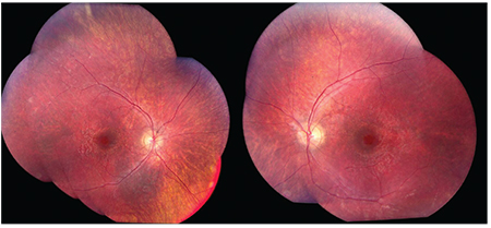 |
| Figure 1. Montage photos of the fundus and mid-peripheral retina depicting blunted foveal reflex, a pseudo “bulls-eye” appearance of the macula, in addition to peripheral pigmentary mottling noted by the examiner. |
Ocular examination revealed a best-corrected visual acuity of 20/30 in each eye. His cycloplegic refraction was +2.25 sphere in the right eye and +2.00 sphere in the left eye.
His pupils were equal, round and reactive without an afferent pupillary defect or paradoxical pupil. His extraocular motility was full and there was no nystagmus. His intraocular pressure by Goldmann applanation tonometry was 10 mmHg in both eyes. Both external examination and slit-lamp examination of the anterior segment were normal. Dilated fundus examination revealed healthy optic nerves, but demonstrated a blunted foveal reflex with a well-circumscribed area of macular pigmentary mottling that resembled a pseudo “bull’s-eye” appearance (See Figure 1). Additionally, wrinkling irregularities of the internal limiting membrane were noted in the macula of both eyes.
In the mid-peripheral retina, there was pigmentary mottling without spicules or clumping of pigment. The retinal vessels were mildly attenuated bilaterally.
What is your differential diagnosis? What further workup would you pursue?
Please click this link for diagnosis, workup, treatment and discussion.


