A 2-year-old Caucasian female presented for evaluation of increased pigmentation of her right iris. This was associated with nodular lesions in the same eye. Per the patient’s parents, the darker iris color and iris lesions had been present since birth. However, the iris lesions appeared to be enlarging in size. On review of systems, there was no history of trauma, weight loss, poor feeding, abnormal bowel or bladder function, fever, rash or recent illness.
Medical History
The patient was otherwise healthy and with normal development. There was no relevant medical, ocular or surgical history. She was not taking medications. Family history was noncontributory.
Examination
Vital signs were within normal limits. Ocular examination demonstrated a visual acuity of fix and follow OU. Pupils were equal and reactive to light without a relative afferent pupillary defect. Extraocular motility was full bilaterally and intraocular pressures were normal by finger tension.
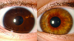
External examination revealed normal eyelids with no evidence of pigmentation, edema or ptosis. Heterochromia was detected with a dark brown right iris and light brown left iris (See Figure 1). The left eye was normal anteriorly and funduscopically.
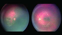
Evaluation of the right eye disclosed diffuse ocular melanocytosis involving the sclera and iris, but sparing the 1 o’clock meridian. The iris crypts were camouflaged with pigment. Additionally, there were multiple, large iris pigment epithelium (IPE) cysts at the pupillary margin and within the mid-zonal region. Funduscopically, diffuse choroidal melanocytosis was noted, with sparing from 10 o’clock to 2 o’clock (See Figure 2). There was no evidence of tumor, orange pigment or subretinal fluid. Fluorescein angiography demonstrated a mildly “silent” choroid in the right eye from the extensive pigmentation.
|
Please click this link for diagnosis, workup, treatment and discussion.


