Presentation
A 27-year-old African-American male presented to the Wills Eye Emergency Room with the chief concern of blurry vision in both eyes. He had been in a motor vehicle accident two weeks prior and since then had been experiencing visual symptoms. He initially noticed flashes of light and floaters with a “blurry smudge” in the periphery of his right eye. For the past three days he felt his left eye was developing “blurry spots.” There was no associated ocular pain, irritation or photosensitivity. The patient denied headache or neck pain.
Medical History
The patient was diagnosed with bilateral keratitis by his optometrist one month prior to presentation. He was treated with polymixin B, which was discontinued prior to his visit to the emergency room. His past medical history was also significant for chronic sinusitis. He was not taking any chronic medications.
His family history was significant for diabetes and cerebrovascular disease. The patient self-identified as a sexually active homosexual who did not consistently use protection. He worked as a nurse and was up-to-date on vaccinations. He smoked tobacco, but denied alcohol and intravenous drug use.
His review of systems was positive for a non-tender, non-pruritic red rash on his chest and abdomen that had been present for “a few weeks.” He also noted right wrist and ankle pain as well as fatigue. He denied fever, chills, night sweats, weight loss, nausea and vomiting.
| ||||
The patient’s ocular examination revealed a corrected visual acuity of 20/25 in each eye. Color plates were 10 out of 10 briskly in each eye. Gross exam showed no mass or proptosis. Pupils were briskly reactive without an afferent pupillary defect. The patient had full motility with normal ductions and versions. Visual fields were full by confrontational field testing. Intraocular pressure was 16 mmHg in both eyes.
The anterior slit lamp exam revealed clear corneas with four white blood cells per high-power field in the anterior chamber of both eyes without evidence of flare. Additionally, the anterior vitreous was positive for cell in both eyes.
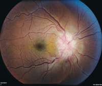
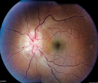
The dilated fundoscopic exam revealed vitreous snowballs in both eyes. There was significant disc edema bilaterally with small, peripapillary cotton wool spots in the left eye (See Figure 1). The periphery was significant for inferior snow-banking and vascular sheathing in both eyes. Further physical examination revealed an erythematous maculopapular rash on the chest and abdomen.
What is your differential diagnosis? What further workup would you pursue?


