A 6-month-old boy of Indian descent was referred from an outside hospital for further evaluation of suspected bilateral aniridia. The patient was initially noted to have an abnormal iris by the pediatrician and referred to a pediatric ophthalmologist, who made a diagnosis of aniridia. The child was referred to a geneticist for appropriate molecular genetic testing. Based on the ophthalmologist’s diagnosis, sequencing of the PAX6 gene was conducted and found to be normal. Concurrent targeted exon-level oligoarray comparative genomic hybridization (ExonArrayDx, Gene Dx, Gaithersburg, Md.) did not reveal a deletion or duplication involving the PAX6, DCDC1, ELP4 or WT1 genes. Approximately 80 percent of cases of aniridia can be explained by mutations in PAX6.1 The absence of a deletion in the 11p13 region eliminates the possibility of WAGR (Wilms tumor, Aniridia, Genital malformations, Retardation) syndrome. Therefore, the child was referred to us to clarify the diagnosis.
Medical History
The boy was the product of a single gestation born at 40 weeks, by normal vaginal delivery. He met all appropriate milestones and was noted to be developmentally normal, making eye contact, smiling, laughing, tracking, rolling and sitting up without support. He was on no chronic medications. The maternal side of the family is North Indian, and the paternal side of the family is South Indian. Per report, the maternal grandmother carried a diagnosis of coloboma since childhood.
Examination
The boy’s vital signs, height and weight were within normal limits. Systemic physical exam was remarkable for a healthy young boy of Indian descent with no significant external pathology and normal external genitalia.
Ophthalmologic examination revealed normal visual responses for age. Intraocular pressures were 8 mmHg OU by Icare tonometry. Pupils were fixed, round and dilated to 8 mm, with a normal margin demonstrating a physiologic rim of posterior pigmented iris epithelium. The iris was symmetric for 360 degrees in each eye and between eyes (See Figure 1). There was no change in pupil size after instillation of phenylephrine 2.5% and cyclopentolate 1%. Eye movements were full and there was no strabismus or nystagmus. Adnexa, lids and lashes were normal. The conjunctivae were white and quiet. The corneas were clear with no limbal pannus. Slit-lamp examination revealed tiny persistent pupillary membrane strands arising from a poorly developed collarette. Anterior chambers were deep and quiet, and lenses were central and clear with mild residual anterior peripheral tunica vasculosa lentis. Fundus examination was entirely normal OU with well-developed fovea, macula and optic nerves. Cycloplegic refraction was OD +8.00 sphere and OS +7.50 sphere.
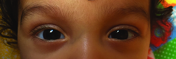 |
| Figure 1. Photograph of the eyes is notable for small, 1 to 2 mm iris remnants spanning 360 degrees with large pupils in the undilated state OU, unchanged post-dilation. |
What is your differential diagnosis? What further workup would you pursue?
Please click this link for diagnosis, workup, treatment and discussion.
Please click this link for diagnosis, workup, treatment and discussion.


