Andover, Mass.
The introduction of Allergan's Botox in 1991 started a boom in the cosmeceutical industry. Since then, the market for cosmeceuticals has steadily expanded, with the introduction of Allergan's eyelash-enhancing Latisse being the latest example, as consumers constantly demand products to alter their cosmetic appearance, since they find over-the-counter cover-ups to be inadequate. One of the primary targets of both marketers and patients alike is the aging eye, specifically the area around the eye. The cosmetics counter offers products aimed at correcting or hiding eyelid swelling caused by age, diet, salt intake, sleep and even the time of day. As no one wants to have an ungracefully aged or unhealthy appearance, products such as Garnier Nutritioniste anti-puff eye roller gel, enriched with caffeine and pro-vitamin B5, readily attract the consumer's attention.
This lid swelling, or morning eye congestion, can be largely attributed to two factors: natural, age-related changes in the lid tissues; and sub-acute localized inflammation that may be triggered by the presence of pro-inflammatory tear film mediators in the closed-eye tear film. While lid swelling and conjunctival redness are not typically sight threatening, the ever-increasing popularity of redness relievers, topical creams and surgical procedures suggest that such quality-of-life considerations warrant clinicians' attention to MEC.
The Sleeping Eye
When we close our eyes to sleep, the levator palpebrae muscle relaxes as the orbicularis oculi muscle contracts to close the eye and keep it shut.1 During sleep, the preocular tear film shifts from a dynamic, reflex, tear-rich layer to a stagnant, secretory and IgA-rich layer.2-6 Open-eye and reflex tears are constantly replenished as the pumping action of the blink flushes away both microorganisms and inflammatory mediators. Upon eye closure, however, the blink ceases, though the exchange of fresh secretions for surplus tears continues.7 The temporal changes between reflex, open-eye and closed-eye tears exhibit markedly different profiles. Open-eye and reflex tears, for example, contain much more lysozyme, lactoferrin and lipocalin, along with minimal secretory immunoglobulin A. The closed-eye tear, on the other hand, is up to 80 percent sIgA.8
Upon eye closure, alterations to the tear film's components, known as complement conversion, also induce polymorphonuclear (PMN) cell recruitment. This shift has been suggested to be a proactive modification to tackle the microorganisms trapped by the eyelid.7 Most of the complement components of closed-eye tears are 0.5 percent to 2.2 percent of their levels in plasma, suggesting they could derive from plasma leaking through the conjunctival blood vessels during sleep. Exceptions to this are C3 and factor B, which may be released by PMNs or synthesized locally. It is due to the massive presence of PMN cells and sIgA that the closed-eye tear film is defined as being in a state of sub-acute inflammation.8 This accumulation of inflammatory factors results in a stagnant, concentrated tear layer.
Unchecked, these adjustments could induce autolytic damage. Therefore, complement inhibitors are also enlisted. Perhaps locally synthesized, vitronectin is an important complement regulator. Through tear screening, it has been shown that vitronectin progressively increases from reflex (0.08 µg/mL) to open-eye (0.75 µg/mL) to closed-eye tears (3.65 µg/mL).7 Vitronectin can potentiate phagocytosis by PMN cells, enhance the processing of trapped microorganisms, regulate the complement proteins through controlling the fate of membrane attach complex and restrict plasmin activation. The ability of vitronectin to bind to partially formed MAC and terminate its completion is its most direct mode of action for complement regulation.7
Nightly phagocytosis can be thought of in terms of minor infection or inflammation. Many of the cardinal signs of infection are present, whether caused by periorbital bacteria, bacteria trapped on the "collecting plate" of the ocular surface or contaminated makeup. Complement conversion leads to an increased presence of inflammatory mediators. These mediators then recruit neutrophils, with maximum recruitment observed after eight hours of sleep.9 As the neutrophils phagocytose pathogens, and subsequently die, both become major constituents of the pus-like discharge sealing the eye upon awakening. In a well-controlled study of non-contact lens wearers, peak redness, temperature and blood flow were documented when measured at waking, with diurnal variations throughout the day.10
The Aging Eye
"Puffy" lids are a frequent cosmetic complaint of aging individuals, especially women. Although many creams and homeopathic remedies purport to treat this ailment, understanding the underlying biological processes sheds light on the inherent shortcomings in these methods. The skin surrounding the eye is thinner than any other on the body, and becomes even more so with sun exposure.11 Elasticity of the skin also decreases with age,11 with the compound effect that the eyelids begin to droop and sag with time. The orbital septum, acting as a girdle, forms the anterior boundary of the orbit.
It, too, weakens with age as collagen and elastin break down, eventually allowing infraorbital fat pads to herniate forward. Also a product of collagen breakdown, tissue turgor, or the ability of the skin to spring back into shape, is lost around this time.
The extracellular matrix is composed of three types of fiber: collagen; reticular and elastic. Collagen fibers, the largest of the three types, consist of thin fibrils tightly compacted to form cord-like shapes.12 These are made up of three intertwined polypeptide chains called tropocollagen bundles. Collagen fibers support the framework of tissues and cells, and are connected to the basal laminae of epithelial cells by reticular fibers. Reticular fibers are more delicate and are made of type-III collagen. These fine fibrils can be individualized or grouped but are continuous with collagen fibers.12 Together, they cross-link to form a meshwork. The elastic fibers are the smallest and typically aren't packed together; individual fibers may be present or bundles may form. The elastin system acts as a buoy to homogeneously distribute stress and establish tissue resilience.12 During the aging process, all three fiber-types relax to some extent. The reticular fibers may have a larger impact on swelling because they provide the space for extracellular fluid.12
Skin seems to thin more in postmenopausal women than in aging men. The cells of the adipose tissue, the major site of extraglandular estrogen production, are some of the cells supported by the reticular fiber system,12 which ensheathes them. Collagen and reticular fibers degenerate and become disorganized, causing them to lose rigidity. Once the elastin network degenerates, elasticity is also lost.13 These fibers fray and allow for adipose herniation.
Hyaluronic acid is a naturally occurring polysaccharide found in the dermis. Due to its affinity for water, it is responsible for providing tissue turgor.14 Turgor can be tested with a simple pinch test: If the skin quickly snaps back into place, tissue turgor is well maintained; if it slowly slides back, turgor is poor. Turgor pressure is the result of the outward force exerted by cell contents on their membrane. Without it, the skin becomes flaccid, and fluid can easily drain from the blood vessels to the eyelid tissue due to decreased extracellular pressure. This interstitial fluid then pools and presents as swelling.
Clinical Considerations
The phenomena associated with the aging lid are exacerbated by several factors. The inflammatory mediators of the closed-eye tear film lead to a state of sub-acute inflammation. The horizontal sleep position, both prone and supine, then allows for fluid drainage into the lower eyelid. Prolonged exposure to sunlight without UV protection and other harmful environmental stimuli such as smoke and pollution also aggravate the situation; even an individual's diet can have a profound effect. Consuming large amounts of salt can lead to dehydration and decrease in tissue turgor.
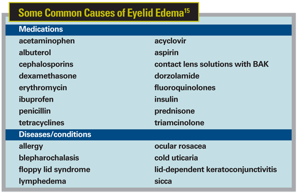
As eyelid swelling can be the result of many etiologies,15 classical signs of other diseases should be kept in mind to prevent misdiagnosis. Because many medications can cause eyelid swelling, a two-week moratorium on corticosteroids, acetaminophen, aspirin, non-steroidal anti-inflammatories and hormonal supplements can rule out a medicinal cause.15 Patient histories are also very important. Through these, MEC can be distinguished from allergy by documented systemic symptoms, and the slow, natural progression of aging can be observed. If a patient complains of "rapid aging," something else may be the culprit. MEC may be present in people of all ages but is increasingly prevalent with age. The signs—morning ocular redness and morning eyelid swelling—are hallmarks of many diseases. The difference, in MEC, is that they are persistent and do not always co-manifest with systemic symptoms.15
MEC is characterized by significant redness and lid swelling that decreases as the day progresses. It's not progressive and there is no pain, heat or lid redness associated with it. Furthermore, it doesn't impair vision or cause diplopia. It is not associated with an S sign, a hallmark of orbital cellulitus in which the closed lid is curved in an S-like shape, and there is no proptosis.
Treatment Options
Current therapy for MEC falls short of user expectations. Makeup can only begin to hide imperfections, and it tends to cake in skin folds and can aggravate the situation if it is contaminated. Creams and other topical applications actually work backwards; they aim to reduce swelling from the outside in rather than inside out, the way eyelid swelling manifests. Creams may have a preventive effect in that they are designed to preserve the health of the skin, but this provides no relief to individuals already suffering from lost tissue turgor and MEC.
While MEC, and more specifically lid swelling, will eventually go away as a result of gravity, it may take hours, leading to quality of life implications. Over the course of the day, skin thickness has been demonstrated to decrease in the upper half of the body while simultaneously increasing in the lower half. This suggests that gravity is able to shift dermal fluid from the face to the legs.16 By this method, eyelid edema can slowly decrease as the interstitial fluid drains. Clinical experience has shown that the swelling can still last up to six hours unabated. It would be more appropriate to treat lid swelling by pulling the fluid from the swollen lid at the location of its origin.
MEC is eventually remedied when the upright, waking position and blinking are re-assumed. However, MEC's effects can last for hours and are hardly affected by homeopathic or natural remedies or cosmetics. Thus, a hole exists in the rising cosmeceutical market: a proven treatment for the signs and symptoms of MEC.
Dr. Abelson, an associate clinical professor of ophthalmology at
1. Franzco AAM. The eye and sleep. Clin Exper Ophth 2005;33:117-25.
2. Tan KO, Sack RA, Holden BA, Swarbrick HA. Temporal sequence of changes in tear film composition during sleep. Curr Eye Res 1993;12:11:1001-7.
3. Sack RA, Tan KO, Tan A. Diurnal tear cycle: Evidence for a nocturnal inflammatory constitutive tear fluid. Invest Ophth Vis Sci 1992;33:3:626-40.
4. Sack RA, Conradi L, Krumholz D, et al. Membrane array characterization of 80 chemokines, cytokines, and growth factors in open- and closed-eye tears: Angiogenin and other defense system constituents. Invest Ophth Vis Sci 2004;45:4:1228-38.
5. Sack RA, Conradi L, Beaton A, et al. Antibody array characterization of inflammatory mediators in allergic and normal tears in the open and closed eye environments. Exper Eye Res 2007;85:528-38.
6. Sack RA, Beaton A, Sathe S, et al. Towards a closed eye model of the pre-ocular tear layer. Prog Retin Eye Res 2000;19:6:649-68.
7. Sack RA, Underwood PA, Tan KO, et al. Vitronectin: Possible contribution to the closed-eye external host defense mechanism. Oc Immunol 1993;1:4:327-36.
8. Willcox MDP, Morris CA, Thakur A, et al. Complement and complement regulatory proteins in human tears. Invest Ophth Vis Sci 1996;38:1:1-8.
9. Thakur A, Willcox MDP, Stapleton F. The proinflammatory cytokines and arachidonic acid metabolites in human overnight tears: Homeostatic mechanisms. J Clin Immunol 1998;18:1:61-70.
10. Duench S, Simpson T, Jones LW, et al. Assessment of variation in bulbar conjunctival redness, temperature, and blood flow. Optom Vis Sci 2007;84:6:511-6.
11. Takema Y, Torimoto Y, Kawai M, Imokawa G. Age-related changes in the elastic properties and thickness of human facial skin. Br J Dermatol 1994;131:641-8.
12. Ushiki T. Collagen fibers, reticular fibers and elastic fibers: A comprehensive understanding from a morphological viewpoint. Arch Histol Cytol 2002;65:2:109-26.
13. Kligman AM, Zheng P,
14. Klein AW, Elson ML. The history of substances for soft tissue augmentation. Dermatol Surg 2000;26:1096-1105.
15. Sami MS, Soparkar CNS, Patrinely JR, Tower RN. Eyelid edema. Semin Plast Surg 2007;21:24-31.
16. Tsukahara K, Takema Y, Moriwaki S, et al. Dermal fluid translocation is an important determinant of the diurnal variation in human skin thickness. Br J Dermatol 2001;145:590-6.


