How do you describe a good tear film? Put simply, a good tear film is sticky on the bottom, juicy in the middle and greasy on top. While this may be an oversimplification, it’s also a good way to start a conversation about mucins—a vital component of a healthy ocular surface. Lipid meibum supplies the greasy top surface that reduces aqueous evaporation, lacrimal glands produce the tears that compose the aqueous layer, and the sticky mucins create an intimate layer of protection at the corneal and conjunctival surfaces. Mucins are tremendously complex molecules in both form and function, and by applying the latest techniques of biological and chemical analysis, we have learned much about their roles in ocular homeostasis and ocular disease.
This month, we review the latest in mucin biology and discuss how modulation of mucins can be used in future treatments of ocular surface disease.
Mucins Explained
Mucins are glycoproteins; they are a protein backbone modified by the covalent addition of multiple, long carbohydrate chains composed of repeating sugar molecules strung end to end. Many include branched carbohydrate chains, adding to the complexity. As with nucleic acids and proteins, this recurring theme of simple units exhaustively combined in seemingly endless chains provides a great opportunity for diversity. So it is with mucins.
There are about 20 basic types of mucins present throughout the body, and of these at least seven or eight have been positively identified at the ocular surface. The varieties of carbohydrate chains and branching patterns within each mucin type also vary, but most range between two and 20 sugar moieties per branch. Despite this heterogeneity, mucins can be categorized functionally as either membrane-anchored or soluble, secreted molecules.
The tethered, membrane-associated mucins are produced by both conjunctival and corneal epithelial cells, and they provide a base coat for the entire ocular surface.1 The polypeptide chains consist of short peptide repeats, including both serine and threonine. These amino acids feature hydroxyl groups on their side chains that provide sites for the O-linkages between protein and sugar chains. Two physical properties are the keys to mucin function: First, as mentioned above, they are sticky; second, this adhesive quality especially applies to “like compounds” such as other mucins.2,3
The primary membrane-associated ocular mucins include MUC1, MUC4 and MUC16.3,4 One important issue that remains unresolved is the relative expression levels of these mucins in conjunctival, corneal or lacrimal epithelium.5-7 Regardless of their tissue locations, these membrane-bound mucins form the base upon which the tear film is built. The adhesive property of mucin molecules is central to their role as physical defenders of the epithelia of all tissues, including the eye’s. In fact, much of what we know about the properties of membrane-associated mucins comes from studies of epithelial-derived tumors, where over-expression of MUC1 and MUC4 plays a role in anchorage-independent growth of some cancerous cells.8
Soluble mucins function in concert with, and independent from, the tethered mucins at the cell surface. Soluble forms are derived from two cell types: conjunctival goblet cells (which primarily produce MUC5AC, also referred to as a “gel-forming” mucin) and the lacrimal acinar cells (which primarily produce MUC7).3,4 Some of these, particularly MUC5AC, associate with membrane-bound mucins to form the mucin layer. Other soluble mucins remain in the bulk aqueous layer and act as lubricants. The stable complexes formed between membrane and soluble mucins create a flexible, protective layer coating the ocular surface. This layer forms a physical barrier to foreign-object access (such as bacteria) to the cellular surface.
The high sugar content of this cell coat gives the layer its second property: It’s hydrophilic, and thus maintains a high water content. This provides a medium through which nutrients, salts and gases (particularly oxygen and carbon dioxide) can pass. This is especially critical for the cornea, an avascular tissue that utilizes adjoining fluids (such as the tear film and the aqueous humor) for its circulatory functions.
Mucins’ Other Functions
We know that mucins are an integral part of the tear film, and we know they serve an important barrier function at the ocular surface. What else do mucins do? Circumstantial evidence suggests that they are part of multiple intracellular signaling pathways.1 There is evidence from a number of studies that mucins function as reporters of tissue damage, eliciting a signal cascade that could lead to epithelial-cell proliferation. They may also serve as tear-film sensors, signaling changes in tear osmolarity.9 It may be that the mucins expressed in the lacrimal glands play a role in this, as they are part of the cells that respond to changes in tear osmolarity. As they do in the conjunctival and corneal epithelium, lacrimal gland epithelia express MUC1 and MUC4, among others.1 Another possible role is suggested by studies of MUC1 splice variants,10 which show a correlation between the relative expression of specific variants in dry-eye patients and the strength of the inflammatory signaling cascade.
One interesting aspect of all of the membrane-bound mucins is that they can, in certain circumstances, be cleaved from their membrane tether and released into the tear film.1 The causes and consequences of this “mucin shedding” aren’t clear, however. The idea that shedding could be a response to bacterial cell binding to the mucin layer, while attractive, has not been demonstrated to date.
Do soluble or gel-forming mucins function as signaling molecules? While we don’t have examples of specific signaling pathways impacted by these mucins, we do know they act both passively and actively as defenders of the ocular surface. An intruding bacterium is coated in mucin, facilitating the process of removal from the ocular surface. In addition, mucins are thought to have antimicrobial activity that further prevents successful colonization of the tear film and its environs.3 It’s important to note that none of the soluble mucins are secreted by corneal epithelia, and so the maintenance of the mucin layer over the entire ocular surface requires the dispersive action of the lid wiper structure of the eyelid margin to distribute mucins across both the conjunctival and corneal surfaces.11
Mucins are also diagnostic tools– when examined carefully, they provide clues to the state of a patient’s ocular surface. White, sticky mucins accompany bacterial infection; fine, stringy mucins are often found in dry-eye sufferers; and a thick, elastic mucin is common in vernal keratoconjunctivitis. Each of these examples shows how mucins respond to different ocular challenges in subtly different ways.
It’s clear that mucins play a host of critical roles as part of the tear film; they are lubricants, anti-bacterials and environmental sensors. They constitute the last line of ocular defense.
Mucin Expression
Several factors regulate mucin expression. Membrane-bound forms seem to display constitutive expression, although the patterns of expression exhibited throughout the course of epithelial-cell maturation change. Soluble mucin biosynthesis, especially the synthesis of MUC5AC, follows with the maturation of the conjunctival goblet cells, which differentiate from progenitor cells scattered throughout the conjunctival epithelium. Mature goblet cells are under parasympathetic control, so drugs with anti-cholinergic side effects elicit dry-eye symptoms, at least in part, by reducing mucin secretion.
There is a host of other factors that also appear to regulate both the maturation of goblet cells and subsequent MUC5AC production; these include serum growth factors such as epidermal growth factor and neurotrophins such as nerve growth factor or brain-derived neurotrophic factor.12 These signaling molecules are likely secreted by the lacrimal glands, while other tear components, including vasoactive intestinal peptide, calcitonin gene-related peptide and neuropeptide Y, are released from nerve cells, both in the cornea and in the lacrimal gland.
All of these neurotransmitters and growth factors have been shown to stimulate MUC5AC synthesis or expression or both, in one or more model systems. In the eye, the best data is from studies of NGF and related neurotrophins.12,13
Mucins and Dry Eye
It’s not too hard to see the importance of a healthy mucin layer for the overall maintenance of an optimal tear film. The number-one dysfunction of tear-film homeostasis is dry-eye disease, and tens of millions of Americans may experience some degree of dry-eye symptoms.14 But what role do mucins play in the etiology of dry eye? The Dry Eye Workshop of 200715 defined two major types of dry eye: aqueous-deficient and evaporative. While a decrease in lacrimal gland secretion can result in reduced secretion of soluble mucins, neither of these classes of dry eye appears to result from a primary defect in secretion of membrane-bound or gel-forming mucins. Disruption of the mucin layer may be more a product of the dry-eye condition, and a sequela to reduction in the aqueous tear volume, inflammation of epithelial cells, or both.
Several studies have suggested that the tear hyperosmolarity that results from either decreased tear production or increased evaporation may alter the mucin layer, and, in doing so, exacerbate dry-eye symptoms.16,17 However, these studies were of in vitro models, and to date there is no evidence that osmolarity itself can disrupt mucin function. In contrast, a recent study in humans showed that hyperosmolarity had no effect on the survival of conjunctival goblet cells.18 Despite these results, animal models of dry eye that employ chronic exposure to the anti-cholinergic agent scopolamine to reduce goblet cell secretion of MUC5AC do seem to provide a model with many of the signs and symptoms of dry eye in humans.19 Studies that combine this model with a controlled adverse environment20 should help to unravel the complex role of mucins in dry eye. Regardless of the mechanisms involved, it’s clear that patients with dry-eye disease often exhibit altered mucin layer function as part of the overall spectrum of their disease. Fortunately, there are a number of potential treatments on the horizon.
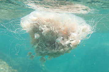 |
| In an innovative move, jellyfish are being studied as a potential source of synthetic mucin for therapeutic use. |
As discussed earlier, mucins are complex molecules composed of many branched-chain carbohydrate monomers. This complexity makes the commercial production of synthetic mucins impractical. Despite this, a recent report21 suggests that a commercially viable source may be in our future: The giant Nomura’s Jellyfish, a species that can grow as long as 12 meters, was used as a source of mucin for treatment of osteoporosis in a recent study. While this treatment is still in the early stages, other therapies are closer to the clinic.
Most mucin-based therapies have focused on enhancing endogenous goblet-cell secretion. Some compounds that have proved effective for treatment of gastric ulcer or irritable bowel disease have been hypothesized to act, at least in part, by stimulation of mucin secretion. Drugs such as lubiprostone or rebamipide are in clinical trials for use as mucin secretagogues in a number of different disease states, including dry eye. Other chemical entities, including flavonoids22 and NGF mimetics,23 have shown promise in preclinical studies. While the complexities of dry eye make clinical development challenging, it’s likely that one or more drugs targeting mucin secretion will make it to market in the near future. Considering mucins’ role in the maintenance of the ocular surface, targeting them seems like a great place to start in building a healthy tear film.
Dr. Abelson, a clinical professor of ophthalmology at Harvard Medical School, consults in ophthalmic pharmaceuticals. Dr. Dartt is a senior scientist and the Harold F. Johnson Research Scholar at the Schepens Eye Research Institute/Massachusetts Eye and Ear, and Associate Professor of Ophthalmology at Harvard Medical School. Dr. McLaughlin is a medical writer at Ora Inc., in Andover.
1. Govindarajan B, Gipson IK. Membrane–tethered mucins have multiple functions on the ocular surface. Exp Eye Res 2010;90:655-663.
2. Guzman-Aranguez A, Argueso P. Structure and biological roles of mucin-type O-glycans at the ocular surface. Ocul Surf 2010;8:8-17.
3. Mantelli F, Argueso P. Functions of ocular surface mucins in health and disease. Curr Opin Allergy Clin Immunol 2008;8:477.
4. Gipson IK. Distribution of mucins at the ocular surface. Exp Eye Res 2004;78:379-388.
5. Barbaro V, Ferrari S, Fasolo A, et al Evaluation of ocular surface disorders: a new diagnostic tool based on impression cytology and confocal laser scanning microscopy. Br J Ophthalmol 2010;94:926-932.
6. Mantelli F, Argueso P. MUC1 biosynthesis in human corneal and conjunctival epithelia. Br J Ophthalmol 2010;94:956-957.
7. Hori Y, Spurr-Michaud S, Russo CL, Argüeso P, Gipson IK. Differential regulation of membrane-associated mucins in the human ocular surface epithelium. Invest Ophthalmol Vis Sci 2004;45:114-22.
8. Workman HC, Miller JK, Ingalla EQ, et al. The membrane mucin MUC4 is elevated in breast tumor lymph node metastases relative to matched primary tumors and confers aggressive properties to breast cancer cells. Breast Cancer Res 2009;11:R70.
9. de Nadal E, Real FX, Posas F. Mucins, osmosensors in eukaryotic cells? Trends Cell Biol 2007;17:571-574.
10. Imbert-Fernandez Y, Radde BN, Teng Y, Young WW Jr, Hu C, Klinge CM. MUC1/A and MUC1/B splice variants differentially regulate inflammatory cytokine expression. Exp Eye Res 2011;Aug 16. [Epub ahead of print].
11. Knop E, Knop N, Zhivov A, et al. The lid wiper and muco-cutaneous junction anatomy of the human eyelid margins: An in vivo confocal and histological study. J. Anat 2011;218:449–461.
12. Rios JD, Ghinelli E, Gu J, et al. Role of Neurotrophins and Neurotrophin Receptors in Rat Conjunctival Goblet Cell Secretion and Proliferation. Invest Ophthalmol Vis Sci 2007;48:1543–1551.
13. Lambiase A, Micera A, Pellegrini G, et al. In Vitro Evidence of Nerve Growth Factor Effects on Human Conjunctival Epithelial Cell Differentiation and Mucin Gene Expression. Invest Ophthalmol Vis Sci 2009;50:4622–4630.
14. Hirsch JD. Considerations in the pharmacoeconomics of dry eye. Manag Care 2003;12:33–38.
15. The Definition and Classification of Dry Eye Disease:Report of the Definition and Classification Subcommittee of the International Dry Eye WorkShop (2007) The Ocular Surface 2007;5:75-92.
16. Li DQ, Chen Z, Song XJ, et al. Stimulation of matrix metalloproteinases by hyperosmolarity via a JNK pathway in human corneal epithelial cells. Invest Ophthalmol Vis Sci 2004;45:4302-11.
17. Luo L, Li DQ, Corrales RM, Pflugfelder SC. Hyperosmolar saline is a proinflammatory stress on the mouse ocular surface. Eye Contact Lens 2005;31:186-93.
18. Moore JE, Vasey GT, Dartt DA, et al. Effect of Tear Hyperosmolarity and Signs of Clinical Ocular Surface Pathology upon Conjunctival Goblet Cell Function in the Human Ocular Surface. Invest Ophthalmol Vis Sci 2011;52:6174–6180.
19. Viau S, Maire MA, Pasquis B. Time course of ocular surface and lacrimal gland changes in a new scopolamine-induced dry eye model. Graefes Arch. Clin. Exp. Ophthalmol 2008;246:857–867.
20. Abelson MB, Ousler GW 3rd, Maffei C. Dry eye in 2008. Curr Opin Ophthalmol 2009;20:4:282-6.
21. Ohta N, Sato M, Ushida K, et al. Jellyfish mucin may have potential disease-modifying effects. BMC Biotechnol 2009;9:98.
22. Choi SM, Seo MJ, Lee YG, et al. Effects of DA-6034, a flavonoid derivative, on mucin-like glycoprotein and ocular surface integrity in a rabbit model. Arzneimittelforrschung 2009;59:498-503.
23. Jain P, Li R, Lama T, Saragovi HU, et al. An NGF mimetic, MIM-D3, stimulates conjunctival cell glycoconjugate secretion and demonstrates therapeutic efficacy in a rat model of dry eye. Exp Eye Res 2011;Jun 25. [Epub ahead of print]


