Today, new technologies may surmount both of those limitations. The latest virtual reality headsets, such as the Oculus Rift (Oculus, Menlo Park, Calif.), allow the visual to follow our changing gaze so quickly that the brain can’t detect a delay. Hence, no more feeling ill. And thanks to the ever-dropping cost of technology, the Oculus Rift will soon be available to the public for a cost of about $300. (Software developers can currently buy a kit to create one for $350.)
This technology could have a drastic impact on many fields, from entertainment (virtual movies in which you can look around during the action) to extremely realistic training simulators (without most of the physical equipment) to mini-vacations (spend a few hours on a tropical island without leaving your home) to 3-D remote interactions with other people.
Here, five individuals using this technology in ways relating to medicine talk about how it’s likely to impact the field of ophthalmology.
Real-world Visual Testing
 |
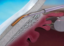
| Using virtual reality to cause a subject to correct for perceived motion has revealed that glaucoma patients’ reactions are more erratic than those of healthy individuals. (Image courtesy Felipe A. Medeiros, MD, PhD.) |
“In real life we use vision in a dynamic way, involving complex scenes and motion,” Dr. Medeiros explains. “The tests we commonly perform to evaluate visual loss in glaucoma are static tests, like standard perimetry fields. We hypothesized that using dynamic stimuli would be a more effective way of evaluating how impaired a glaucoma patient has become.
“We used an immersive 3-D virtual-reality environment to help us evaluate balance in patients who have glaucoma, in comparison to controls,” he continues. “The subjects wore an Oculus Rift while standing on a force platform. We projected a series of different visual stimuli, such as moving through a tunnel (translational stimulus) and standing in a rotating environment (rotational stimulus). Subjects reacted to the perceived movement by shifting their bodies to compensate. The platform measured the force their bodies applied when they attempted to regain or prevent their perceived loss of balance.
“In patients with glaucoma, the responses were not appropriate,” he says. “Their responses were more erratic than those of healthy subjects, and they lost their balance much more easily. More importantly, we found that the metrics produced by this test were predictive of the risk of falling. A history of falls in these subjects revealed that those who had more falls performed worse in the test.” (Potentially being able to predict falling is a big deal; falling is the leading cause of injury-related death in older adults, especially those with glaucoma.)
Dr. Medeiros points out that virtual reality offers ophthalmology a chance to break free of the traditional constraints of in-office testing. “Studies have shown that tests like the standard visual field are not very predictive of how patients actually perform in the real world,” he says. “I see virtual reality as a way of testing things in a more realistic way that will allow us to assess how visual impairment affects our patients’ ability to do things. For example, we have a very elaborate driving simulator in my lab; it’s a full car, so the test is very realistic. With virtual reality we might be able to recreate a portable version of that test.”
Dr. Medeiros notes that the technology involved in the balance test is rapidly becoming less expensive. “An Oculus Rift can be purchased for a few hundred dollars,” he says. “And although we used an expensive force platform in our experiment to maximize our accuracy and the value of our data, the same test might be accomplished using the kind of floor mat people stand on when they play some Wii video games. We’re now working to develop a simplified version of the test that could be routinely used in clinical practice to predict which patients are at higher risk of falling. If you know your patient is at higher risk, you can provide some counseling and perhaps have the patient go into a program designed to reinforce balance through exercise or other means.”
Dr. Medeiros notes that his group’s intention was not to create a diagnostic screening tool. “However, we did find that this test is able to predict the risk of falling even in those with only mild or moderate glaucoma, who may not realize they are at risk,” he says. “It would be interesting to evaluate the potential of this kind of test as a screening tool.”
Another Virtual Vection Test
Researchers at the Vision Science Research Program of the Toronto Western Research Institute are also investigating the use of virtual reality to create vection—the sensation that you are moving—as a way to evaluate the status of glaucoma in a patient. Their results indicate that even individuals with early glaucoma experience vection differently than healthy controls, raising the possibility of a simple screening test for the disease. (They conducted their initial tests seating subjects in front of large screens to create images that fill the entire visual field, but they plan to replace those screens with a virtual reality headset.)
“Our test presents the study participants with a random dot pattern that fills their entire visual field,” explains Taylor Brin, a graduate student in the Ocular Motor Laboratory, which is part of the VSRP. “The dot pattern rotates in a circle. This causes the participant to feel as if he or she is moving; in other words, it induces vection. The subjects simply press and hold a button as soon as they feel like they’re moving. We compared the vection response in individuals with healthy vision to the response from those with mild-stage glaucoma; we found that individuals with glaucoma had vection that was either impaired or completely absent compared to those with healthy vision.”
Ms. Brin says the idea for this test originated when other members of the lab noted studies showing that vection was altered in individuals with age-related macular degeneration. “Initially we did our vection test in macular degeneration patients; we found they have a stronger sensation of vection because they have damaged central vision. That led us to think, why not try this in glaucoma where the damage is sort of the opposite of macular degeneration? Instead of having damaged central vision, people with glaucoma typically have damaged peripheral vision at first.”
Ms. Brin says the early results have been highly statistically significant. “As we hypothesized, individuals with glaucoma have a weaker sensation of vection,” she says. “They either don’t experience vection at all—in contrast to healthy controls who universally experience vection—or it takes them a lot longer to experience it than controls. Notably, we only tested patients with early glaucoma, which makes this very promising as a screening tool. Even patients with 20/20 vision and very few visual deficits showed this effect.”
Ms. Brin says the team is very interested in trying the same test using a tool such as the Oculus Rift. “Reality-based tests involving things like vection are very promising, but performing the test with a large screen is inconvenient,” she notes. “Virtual reality is compact and convenient; it’s easy to have in the doctor’s office, and it’s relatively inexpensive compared to some of the tests we have now. Using virtual reality to perform this sort of test could help to make diagnosis much easier.”
Making Standard Tests Portable
One of the advantages of current virtual reality technology is its portability. Thus, existing tests such as perimetry can be recreated in virtual reality, eliminating the need for bulky, non-portable equipment. Along those lines, CREWT Medical Systems in Tokyo has developed a head-mounted device that can conduct a standard perimetry test without the usual equipment. The virtual test should be available for purchase in Japan later this year.
Shinji Kimura, director and general manager at CREWT Medical Systems in Tokyo, notes that it’s not practical for most surgeons to increase the number of measurements taken in the office. “We chose to develop the head-mounted perimeter partly because its high portability helps to solve this problem,” he says. “In addition, the problem of finding a suitably dark place to give the test is settled because the head mount provides its own darkroom, and finding space for the equipment is no longer an issue.
“This device is equipped with an optical system that provides a field of vision of 35 degrees from the center to the right and left,” he continues. “It uses a liquid crystal display and can create the broad dynamic range of brightness and various sizes of stimulus necessary for a perimeter. It makes it possible to measure perimetry binocularly, and there’s no need to close one eye for a monocular test. In fact, when performing binocular random perimetry, it is impossible for the subject to know which eye is being tested. The device can also monitor the pupil with high precision and use that information to control the stimulus point, making it easy for the subject to maintain fixation.” Mr. Kimura notes that the device is balanced to avoid feeling too heavy on the head, although he admits some elderly individuals might be bothered by the weight.
Mr. Kimura says they have conducted a study comparing the device to a standard perimeter. “The correlation is very good, but the study has not yet been published, so I can’t share the numbers,” he says. He adds that the company is currently conducting a study including a questionnaire to help determine how subjects feel about taking the test in this way. They hope to eventually bring the product to the United States.
Dr. Medeiros, along with members of the neuroscience department at the University of Southern California, San Diego, is also working on a virtual reality-based perimeter that takes things one step further: It removes the subjective part of the test by measuring visual responses using electroencephalograph technology embedded in the headset worn by the patient. “This not only eliminates the need for the standard perimeter, it will be an objective test of visual impairment that does not rely on subjective patient responses,” he says. “We’ll analyze the patient’s brain waves to determine whether he saw the stimulus or not. We’ve already created a prototype of the device.”
Capturing Virtual Live Surgery
The MOVEO Foundation, based in France, focuses on research projects aiming to modernize orthopedic surgery. Recently, the Moveo Foundation supported a project that pioneered the capturing of surgical procedures in 3-D; the procedures are then translated into a virtual reality experience that can be shared via the Oculus Rift, for multiple purposes including training.
 |
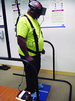
| France’s Moveo Foundation has funded 3-D, open-field recording of surgeries (note cameras attached to surgeon’s head), which are then translated into virtual reality format. The viewer can turn and look in any direction during the virtual playback. (Image courtesy Julie Mercier/Moveo Foundation.) |
“When you watch the procedure through the virtual reality headset, you can move your head in this virtual world,” he continues. “If you are more interested in what the assistant is doing, or the scrub nurse, just turn your head toward them to see what they are doing; the stereoscopic 3-D and wide field of view create a striking immersive effect. A student can replay the surgery in detail and watch it through the eyes of the surgeon. Surgeons can also use this to observe and learn from their own work.”
Taking the element of reality one step further, an English product-development firm, Plextek Consultancy, has been working with its government’s Defense Science and Technology Laboratory to create an immersive virtual reality simulation training system for medical members of the military, using the Oculus Rift. The system creates battlefield environments that can be experienced by multiple individuals at once, allowing them to learn to treat casualties in three-dimensional, high-stress, under-fire situations. Notably, the system also records the participants’ actions while responding to the virtual situation for later evaluation.
“We’ve enabled the system to record everything that happens, monitoring the actions of the participants, visually and verbally,” explains Collette Johnson, medical business development manager at Plextek. “Everything is time-stamped and searchable. This should be very useful for surgeons in training; you can see whether the person was following the steps correctly. It could also be useful for a surgeon who simply wants to review his own surgery. What makes this different from simply filming a procedure is that it’s an intelligent system and a data pool; you can search for key moments and go straight to them; you can search for specific words in the audio track. And of course, because it’s virtual reality, you can look around, 360 degrees, at any moment during the recorded procedure. And it can automatically become part of the electronic record, if desired.”
What Lies Ahead?
“Like other highly demanding surgical disciplines, ophthalmology will benefit from affordable immersive systems such as virtual reality,” says Dr. Gregory. “This will be a powerful way to train young surgeons, especially as the demand for remote training systems increases with the development of health-care systems in emerging countries. The potential of virtual reality is also huge for patient care, as it can be used advantageously in the framework of minimally invasive surgical techniques.”
Ms. Johnson sees multiple potential uses for virtual reality. “Virtual reality can allow patients and family members to experience procedures and their benefits ahead of time,” she notes. “For example, many patients are nervous about getting an injection in the eye. With virtual reality they and any concerned family members can see what’s involved. Virtual reality could also be used to conduct tests that would normally have to be done in the office, tests that can detect musculature problems or neuro-ophthalmological problems, and conduct them outside the office in the community. That might make it easier to discover problems earlier when they’re more amenable to treatment.”
Ms. Johnson also sees virtual reality helping individuals with low vision experience things that would be challenging or impossible to experience firsthand. “An individual with low vision might want to visit the Metropolitan Museum of Art, but find it very frustrating—if he could even get there,” she notes. “Virtual reality would allow that person to spend as much time as desired interacting with the art, as closely as desired, either in the museum or at home. Possibilities like this might help to prevent some of the side effects we see in people with low vision, such as depression and feeling isolated. This technology is already helping people who can’t go outside for other reasons, such as anxiety about being in public, or individuals with late-stage cancer who simply can’t travel.”
Dr. Medeiros also sees great potential in using virtual reality to overlay 3-D data on live images—which he refers to as augmented reality—such as the images seen through a microscope during surgery. “Having a reticle overlaid on the microscope view is a simple version of this,” he says. “I think this will go far beyond that. You could overlay topographic data, or a 3-dimensional rendition of the best incision architecture for a given case. Companies are already developing augmented reality glasses that will allow you to see a patient’s records and data while talking to the patient. You might even be able to turn the page or move the visuals around by waving your hand, as the characters did in the movie ‘Minority Report.’ ” REVIEW
1. Diniz-Filho A, Boer ER, Gracitelli CP, Abe RY, van Driel N, Yang Z, Medeiros FA. Evaluation of Postural Control in Patients with Glaucoma Using a Virtual Reality Environment. Ophthalmology 2015;122:6:1131-8. doi: 10.1016/j.ophtha.2015.02.010. Epub 2015 Apr 16.