The Kamra Candidate
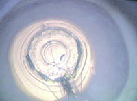
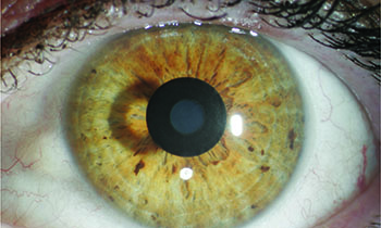
To understand what makes a good Kamra patient, it helps to quickly review the device’s design. The Kamra is a small, thin, ring-shaped inlay 3.8 mm in diameter, 5 µm thick, with a central 1.6-mm aperture. The ring itself is riddled with approximately 8,400 holes that are each 5 to 11 µm wide, which allow the flow of nutrients to the cornea. The Kamra procedure involves the use of a femtosecond laser to create a pocket at a depth of 200 µm in a patient’s non-dominant eye, into which the Kamra is placed using special forceps. Once in place, the Kamra is designed to increase the depth of focus in emmetropic presbyopes through the pinhole effect, while having a minimal effect on distance vision.
Tokyo surgeon Minoru Tomita has implanted just over 10,000 Kamra inlays, and has learned a good deal about how to make them work in patients. “In the United States, the prospective patient’s spherical equivalent refraction should be between -0.75 and +0.5 D,” says Dr. Tomita. “So the candidate pool is very small.” Surgeons outside the United States have learned to expand the candidate pool by combining the Kamra with LASIK, though this isn’t in the official FDA labeling for the device. “I’m combining it with LASIK, so if the patient has hyperopia of +3 D I can make it -0.75 D with a hyperopic LASIK procedure. Similarly, if the patient is -5, I can make him -0.75 with a myopic LASIK,” says Dr. Tomita. Jay Pepose, one of AcuFocus’s clinical investigators in the U.S. trial, says this range of vision is ideal for these patients. “Probably the -0.75 D is really the sweet spot, because you’re extending the depth of field.”
|
Dr. Tomita says that, as in other areas of refractive surgery, a patient who is very nervous or tends to obsess over imperfections may not be a good candidate. “If the patient is very sensitive or nervous, we wouldn’t choose the Kamra for him,” he says. “This is because, sometimes, it takes time to recover the vision with the Kamra inlay. Most patients get their distance and near vision back in a week, but some patients take several weeks to one month. A nervous patient can get very worried about that, and can take a lot of your time having many discussions about it.”
Implantation Pearls
Between the U.S. study and the experience of surgeons outside the United States, physicians have developed certain best practices when approaching a Kamra implantation.
• Laser settings. Though it was initially thought the Kamra would go beneath a LASIK flap, the device’s labeling specifically calls for a femtosecond-created pocket, so the Kamra surgeon needs access to a femtosecond laser. “Prior to the study, many surgeons were using a flap, but this created more dry eye,” says Dr. Pepose. “The pocket is clearly the preferred way to go about implanting it. Also, as we carried out the FDA study of the device, it turned out that the laser settings were critical. A tight line and spot setting, less than or equal to 6 x 6, is important. When we started to go higher in the line/spot separation, the tissue around the pocket that needed to be separated was like Velcro, and you’d encounter areas that weren’t lased in between the spots. This led to the creation of more inflammation as the surgeon dissected the pocket, and these patients would not fare as well as the patients who had the tighter spot. The tighter laser spacing reduces the wound-healing response and enhances refractive stability.”
• Depth. Surgeons say the 200-µm depth is a good spot for avoiding corneal issues. “We didn’t really stratify the data from the study at different depths, but you don’t want to go too shallow,” says Dr. Pepose. “In previous studies, when they went too shallow they had issues with corneal thinning, and you also don’t want issues from the change in shape of the cornea by going too shallow; you don’t want changes in topography. At 200 µm, you’re also not so deep that you have concerns of ectasia.”
• Centration. Though it’s important to have the Kamra centered properly, surgeons say it’s actually very forgiving when it’s not dead center. In addition to a femtosecond laser, Dr. Pepose notes that surgeons will need access to AcuFocus’s AcuTarget HD device to implant the Kamra. The device helps surgeons locate landmarks and Purkinje images to guide Kamra centration. “A lot of the centration depends on the eye’s angle kappa,” Dr. Pepose explains. “Usually, we target the first Purkinje image. If there’s a large angle kappa, then we’ll usually split the difference and aim between the first Purkinje and the center of the pupil.” Surgeons say the inlay can be off by up to 300 µm and still yield good results.
Dr. Tomita says the design of the operating microscope you use during implantation also can make a difference. “You have to make sure that the fixation light strikes the cornea at a 90-degree angle,” he says. “There can’t be an oblique angle from the light to the corneal surface. The Takagi microscope company has a special microscope for the Kamra inlay, and the WaveLight Allegretto 400 and AMO/Visx excimer laser microscopes are usable. You can also use a microscope that you use for cataract surgery. However, you can’t use the Allegretto EX 500 or the iFS microscopes.”
• Don’t fiddle with it. Surgeons say trying too hard to center the device perfectly will backfire. “During implantation, the surgeon, especially the beginner, can be very nervous,” says Dr. Tomita. “In an effort to place it on the center of the Purkinje image, he might try to move it, then find he’s moved it too far, then try to move it back. If you move the Kamra too much, however, patients take more time to recover their vision. If you think it may not be perfectly centered, just wait until the postop period. If the patient doesn’t have good vision, then you can move it. But if you touch the Kamra too many times during the surgery, the patient’s vision will be decreased.”
|
Postop Management
In addition to visual results, surgeons say there are other things to watch for in the postoperative period.
In the pivotal FDA study, at 12 months 84 percent of the patients (399/478) could see 20/40 or better uncorrected at near in the Kamra eye. Binocularly, 94 percent could see 20/40 or better uncorrected at near. In terms of uncorrected distance vision in the implanted eye, patients lost an average of three letters at 12 months.1
Forty-five patients (8.9 percent) had to have their inlays removed over the course of the first five years postop. Two of the removals were for the appearance of the inlay, four removals were medically indicated, and 25 were removed due to visual complaints (25 of these due to hyperopic refractive shifts). All but one of the explanted eyes returned to 20/20 best-corrected distance vision, with the remaining one seeing 20/25, possibly due to a corneal scar. Dr. Pepose says that the explantation rate was higher in the study than it is currently because the study surgeons didn’t yet know about the proper spot/line separation or the optimum preop refraction. “The explantation rate is now around 2 percent in current Kamra usage,” he says.
Surgeons say steroids can help handle some postop situations. “We use a steroid for at least six months to prevent a haze reaction,” says Dr. Tomita. “If they develop haze, patients will get a hyperopic shift—becoming +1 or +2. If this occurs, we’ll use dexamethasone five times daily, and patients will improve after a month or a little longer. After they improve, we’ll reduce the steroid. If there’s no improvement, though, we’ll have to remove the inlay.”
Dr. Pepose says that, if patients know what to expect ahead of time, they can do well with the Kamra. “Like any refractive surgery, you want the patient to have reasonable expectations,” he says. “If the patient wants to be able to take out his smartphone and read it, that’s OK. But, if he wants to sit for five hours and read War and Peace, he may still have to put on low-power readers. The nice thing though, is with the Kamra you’re not changing the second eye’s refraction, so store-bought readers work. In the end, just be sure to establish realistic expectations.” REVIEW
1. AcuFocus Kamra labeling. http://www.acufocus.com/int/sites/default/files/Physician%20Labeling.pdf. Accessed 15 May 2015.


