 |
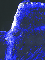
| The white areas in this image show extensive regeneration of nerve fibers (axons) in the central nervous system achieved by gene therapy after optic nerve injury. (The blue areas indicate scar tissue.) (Image courtesy Fengfeng Bei, PhD.) |
As reported online January 14 by the journal Cell, the scientists restored vision in mice with optic nerve injury by using gene therapy to get the nerves to regenerate and—the crucial step—adding a channel-blocking drug to help the nerves conduct impulses from the eye to the brain. In the future, they believe, the same effect could be achieved with drugs alone.
In the study, previously blind mice turned their heads to follow patterns of moving bars after given the treatment, say co-senior investigators Zhigang He, PhD, and Michela Fagiolini, PhD, of the Department of Neurology and F.M. Kirby Neurobiology Center at Boston Children’s. The technicians doing the tests did not know which mice had been treated.
“By making the bars thinner and thinner, we found that the animals could not only see, but they improved significantly in how well they could see,” says Dr. Fagiolini.
While other teams, including one at Boston Children’s, have restored partial vision in mice, they relied on genetic techniques that can only be done in a lab. Generally, their methods involved deleting or blocking tumor suppressor genes, which encourages regeneration but could also promote cancer. The new study is the first to restore vision with an approach that could realistically be used in the clinic, and that does not interfere with tumor suppressor genes.
The key advance in restoring vision was getting the regenerated nerve fibers (axons) to not only form working connections with brain cells, but also to carry impulses (action potentials) all the way from the eye to the brain. The challenge was that the fibers regrow without the insulating sheath known as myelin, which helps propagate nerve signals over long distances.
“We found that the regenerated axons are not myelinated and have very poor conduction—the travel speed is not high enough to support vision,” says He. “We needed some way to overcome this issue.”
Turning to the medical literature, they learned that a potassium channel blocker, 4-aminopyridine (4-AP), helps strengthen nerve signals when myelin is absent. The drug is marketed as AMPYRA for multiple sclerosis, which also involves a loss of myelin. When they added it, the signals were able to go the distance.
While the study used a gene therapy virus called AAV to deliver the growth factors that trigger regeneration (osteopontin, insulin-like growth factor 1 and ciliary neurotrophic factor), He and Fagiolini are testing whether injecting a “cocktail” of growth factor proteins directly into the eye could be equally effective.
“We’re trying to better understand the mechanisms and how often the proteins would have to be injected,” says He. “The gene therapy virus we used is approved for clinical study in eye disease, but a medication would be even better.”
With regeneration kick-started, 4-AP or a similar drug could then be given systemically to maintain nerve conduction. Because 4-AP has potential side effects including seizures if given chronically, He and Fagiolini have begun testing derivatives (not yet FDA-approved) that are potentially safer for long-term use.
The researchers are further testing the mice to better understand the extent of visual recovery and whether their approach might get myelin to regrow over time.
“The drugs might need to be paired with visual training to facilitate recovery,” says Dr. Fagiolini. “But now we have a paradigm to push forward.”
Study Sheds New Light on Keratoconus
A largest-ever clinical study of keratoconus reveals previously unknown risk factors associated with the condition. The new study shows that men, African Americans and Latinos, and people with asthma, sleep apnea or Down syndrome, have much higher odds of developing keratoconus. But females, Asian-Americans and people with diabetes appear to have a lower risk, the analysis shows.
The findings, made by researchers at the University of Michigan Health System’s Kellogg Eye Center and the U-M Institute for Healthcare Policy and Innovation, are published online ahead of print in Ophthalmology.
The research was sparked by questions whether changes to the eye with keratoconus affect other parts of the body. Studying eye conditions’ associations with other health conditions is easier now because of vast data troves.
“Eye health relates to total body health, and we as ophthalmologists need to be aware of more than just eyeballs when we see patients,” says Maria Woodward, MD, an assistant professor of ophthalmology at the U-M Medical School and first author of the new study. The last decade has brought new treatment options, but many people don’t receive a diagnosis early enough to take full advantage of them.
Patients with keratoconus and their families, as well as physicians, should be aware of other potential health problems uncovered in the study, the authors say.
The researchers made their findings by looking at data from health-insurance claims, half of them from more than 16,000 people with confirmed keratoconus and half from an equal number of people with similar characteristics but no keratoconus.
This allowed them to see which characteristics and medical conditions were most associated with keratoconus, and which weren’t. The people in the study were mostly in their 30s and 40s.
The study helps confirm many suspicions about the condition raised by previous small studies, but casts doubt on others. For instance, men were already known to have a higher risk, which the study confirmed.
And people with Down syndrome had a much higher chance of having keratoconus—six times higher than others—a known risk but still a stark one. This reinforces the high importance of screening and treatment for the condition in members of the Down syndrome community, starting at a young age, Dr. Woodward says.
But the higher rates of keratoconus among people of African American and Latino origin—50 percent higher than whites—were previously unknown. And the finding of a 39-percent lower rate among people of Asian heritage contradicts previous research.
Meanwhile, there’s been debate over a possible “protective” effect of diabetes. While diabetes causes other negative effects to the eye, the cornea may be strengthened as a by-product of those changes.
The new finding of a 20-percent lower odds of keratoconus among people with diabetes, and an even lower among those with complications from diabetes, appears to support this idea.
The researchers also looked at other chronic conditions thought to be associated with keratoconus, such as allergic rhinitis, mitral valve prolapse, collagen vascular disease, aortic aneurysm and depression, and found no higher odds of the condition.
But when it came to people who had been diagnosed with sleep apnea, which interrupts breathing during sleep, and can cause snoring, daytime sleepiness and a higher risk of heart disease and stroke, there were statistically significant higher odds of also having keratoconus. Similarly, people with asthma had higher odds of also having the eye condition.
The authors note that because they used insurance data, they can only see associations of conditions recorded on medical bills, and not cause and effect. And, their findings might not apply to people with no health insurance and therefore less access to medical care.
They also can’t tell which of the people had other risk factors for keratoconus, such as eye rubbing, a family history of the condition, and other conditions not present in the database.
Study Confirms Myopia Surge
The largest study of childhood eye diseases ever undertaken in the United States confirms that the incidence of childhood myopia among American children has more than doubled over the last 50 years. The findings echo a troubling trend among adults and children in Asia, where 90 percent or more of the population have been diagnosed with myopia, up from 10 to 20 percent 60 years ago.
MEPEDS, the Multi-Ethnic Pediatric Eye Disease Study, conducted by researchers and clinicians from the USC Eye Institute at Keck Medicine at USC in collaboration with the National Institutes of Health, adds to a growing body of research into the incidence and potential causes of myopia in children and adults.
The possible culprit? Too much “screen time” and not enough sunlight, according to Rohit Varma, MD, MPH, and director of the USC Eye Institute.
“While research shows there is a genetic component, the rapid proliferation of myopia in the matter of a few decades among Asians suggests that close-up work and use of mobile devices and screens on a daily basis, combined with a lack of proper lighting or sunlight, may be the real culprit behind these dramatic increases,” said Dr. Varma. “More research is needed to uncover how these environmental or behavioral factors may affect the development or progression of eye disease.”
The USC study found that the incidence of childhood myopia in the U.S. is greatest in African-American children, followed by Asian-American children, Hispanic/Latino and Non-Hispanic white children. Future research may include re-examining the MEPEDS cohort to evaluate how widespread use of “screens” and other environmental or behavioral factors may be affecting the progression of childhood myopia and other eye diseases over time.
From 2003 through 2011, MEPEDS provided free eye exams at USC Eye Institute clinics to more than 9,000 Los Angeles-area children ages 6 months through 6 years. “In addition to being the largest pediatric eye study ever undertaken, it is the first of its kind to examine children as young as 6 months old,” said Dr. Varma. “Typically, children do not undergo vision testing until they reach school age. By including younger children, we have the opportunity to identify eye diseases and their causes at the formative stages.”
USC Eye Institute researchers and clinicians collected basic health information during a home visit with the child and parents, followed by a detailed eye examination under dilation. REVIEW


