Boston
Optical Coherence Tomography is a non-invasive technology developed more than 15 years ago as an optical biopsy to obtain high-quality, cross-sectional images of biological tissues.1 Since its discovery and integration into clinical medicine, it has become pervasive in the field of ophthalmology, specifically in managing retinal diseases,2 glaucoma,3 and the anterior segment4 via the anterior segment OCT. There are currently two distinct OCT technologies commercially available: the older time domain technology and the newer spectral or Fourier domain OCT technology.
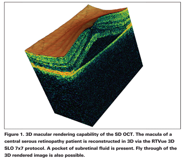
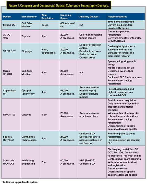
OCTs operate on the principle of indirect interferometry, in which a beam of light is directed into the retina, and the resulting back-scattered light travels an unknown distance to a detector, which is compared to a reference beam of known length to calculate the echo time delay of light. In time domain OCT, the reference arm mechanically moves, and the echo time delays are measured one at a time. In contrast, spectral domain OCTs have a fixed-reference arm to generate an interference pattern of the reflected light, which then undergoes Fourier transformation to measure all the echo time delays simultaneously. The integration of Fourier transformation technology has dramatically increased the image acquisition speed of OCT devices from 400 A-scans/second in the standard time domain Stratus OCT (Carl Zeiss Meditec) to 18,000 to 50,000 A-scans/second in spectral/Fourier domain OCTs. The improved imaging decreases the presence of fixation drift while increasing the total retinal coverage of the OCT scan. The dense scans generated by the spectral domain OCT devices make 3D macular and optic nerve rendering and fly-through possible (See Figure 1).5,6 These functions are now present in the majority of the commercially available devices for qualitative analysis of the retina.
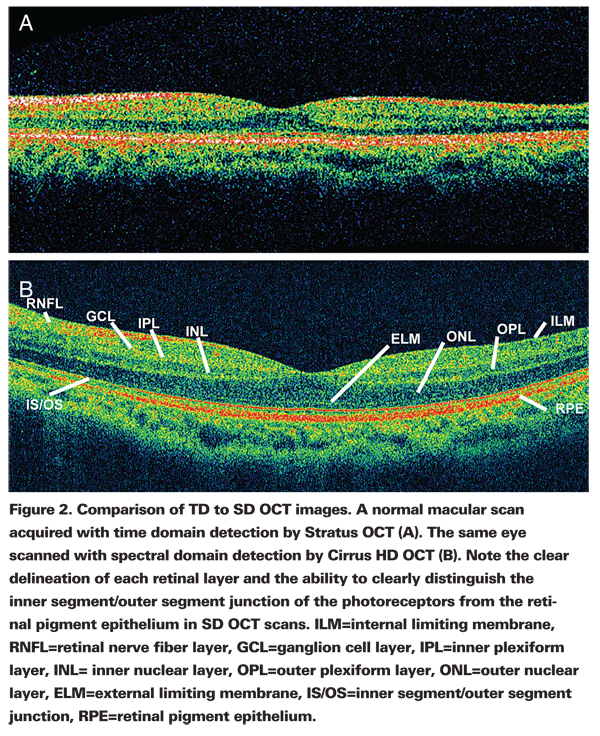
Resolution of OCT scans is inversely proportional to the light bandwidth. Commercial OCT devices utilize low-coherence superluminescent diode (SLD) light sources. Stratus OCT has a bandwidth of about 25 nm centered at 840 nm, giving it a resolution of 10 µm. Commercially available spectral/Fourier domain OCTs have bandwidths near infrared wavelengths, achieving about 3 to 5 µm axial resolution in tissue. The improved resolution allows for clear delineation of each retinal layer as distinct entities, like the ability to differentiate inner segment/outer segment (IS/OS) junction from the retinal pigment epithelium (See Figure 2).
Stratus OCT
The Stratus OCT (Carl Zeiss Meditec) is currently the standard in commercial OCT devices for posterior segment imaging. It utilizes time domain detection and provides realtime cross-sectional images. It has a scanning speed of 400 A-scans/second with a resolution of 10 µm. The main disadvantages of this system are the lower resolution and the slower scanning speed compared to time domain OCT devices. The lower resolution of the Stratus OCT results in no clear delineation of the IS/OS junction from the RPE or other subtle outer retinal pathologies. The slower scan speed means that the device is more prone to motion artifacts due to fixation drift and covers a smaller portion of the retina. Recent study by our group has also shown that Stratus OCT scans contained the highest number of clinically significant segmentation line breakdown (central foveal subfield thickness change by at least 11 µm post-manual correction of segmentation line breakdown) compared to three other spectral domain OCT devices: Cirrus HD OCT, RTVue 100 and Topcon 3D OCT 1000 in a large group of eyes with retinal pathologies (manuscript in submission).7 Time domain OCT scans also revealed lower interscan repeatability compared to scans from spectral domain OCTs.8 However, the Stratus OCT is the most affordable commercial OCT system— it is about 25 to 50 percent less expensive compared to the spectraldomain OCTs. In addition, operators have had the most experience with the Stratus OCT since this device has been around longer compared to spectral-domain devices. Since it is the OCT in widest use, it is also the best-studied device with the most number of published studies. However, it is important to note that studies conducted with time domain OCTs involving measurements of retinal thickness cannot be directly compared to results obtained from spectral-domain devices, since the outer retina is segmented at different locations between time domain and spectral-domain OCT devices.7,8,10 This is a potential hurdle to overcome if the industry transitions from time domain to primarily spectral-domain OCTs.
The macular thickness protocol is the main retinal scan used for this device; it consists of six radial line scans, which are acquired individually. Quick macular thickness protocol acquires all six scans at once but at a lower resolution. The use of the six-line radial scanning pattern requires interpolation of points by the software in the generation of retinal thickness maps. The high dependency on interpolation often leads to loss of important details in these maps, like in the case of fine pathology where the lesion falls between the six intersecting raster lines and is thus missed, leading to inaccurate macular thickness calculation. Denser scan patterns, which require much less interpolation of points, are possible with the high speed of the spectral-domain OCTs, and thus it is now possible to obtain much more accurate depiction of retinal thickness.
3D-OCT 1000
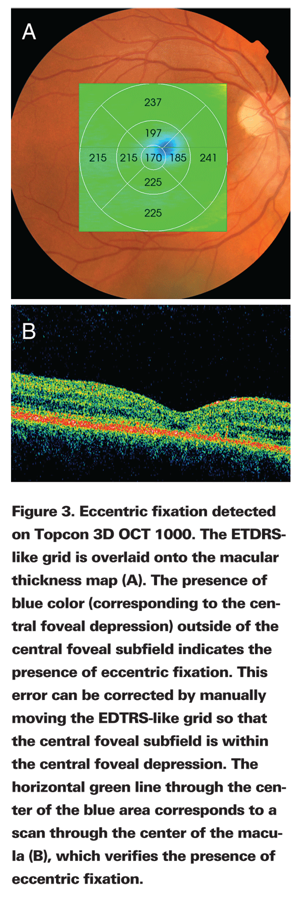
Topcon 3D OCT 1000 (Topcon Medical Systems) has a scanning speed of 20,000 A-scans per second with an axial resolution of 6 µm. The SLD light source used is centered at 850 nm with a bandwidth of 50 nm. The unique feature of this device lies in its integration of a true color non-mydriatic fundus camera, in addition to the spectral-domain OCT. Pinpoint registration of OCT scans is possible by the automatic placement of the OCT projection image (OCT C-scan: enface view of a series of sequential OCT scans down the fundus put together) onto the color fundus image. Besides being a cost and space saving option, this setup makes the correlation of specific lesions observed on the color fundus photo to the corresponding OCT image possible. Various overlays, such as the topographic retinal thickness map and projection image, may be placed over the fundus photo simultaneously with different fundus grids, such as the EDTRS-like map, rectangular map (5 mm x 5 mm map with six boxes horizontally and vertically) and volume map. The superposition of the retinal thickness map with the EDTRS-like map helps to detect eccentric fixation (See Figure 3), which can then be corrected manually. In addition, the 3D OCT 1000 software interfaces smoothly with IMAGEnet and integrates with other types of images like fluorescein and indocyanine green angiography, red-free and autofluorescence and thus it is possible to register fluorescein and other image types and register them to OCT scans.
3D SDOCT
Bioptigen 3D SDOCT is a device clinically suitable for the scanning of patients, in addition to its usefulness in clinical and animal biomedical research. It has a scanning speed of 20,000 A-scans/second and an axial resolution of 5 µm. The device is unique in that it has two light sources, one centered at 1,310 nm and another at 820 nm. The dual light source makes it especially suitable for multiple research settings. The 1,310 nm engine may be clinically helpful for anterior segment imaging and in basic scientific research for tissue, small animal, external and ex-vivo imaging. The 840 nm engine may be clinically helpful for human retinal imaging and in basic science research for retinal imaging in small (i.e., rodents) and large animals (i.e., dogs). A 170-nm bandwidth light source upgrade is also available for high-resolution imaging. Additionally, the Bioptigen 3D SDOCT contains a variety of scanners and probes for various applications such as a Doppler processing system for retinal blood flow analysis, small animal probe for basic scientific research, pediatric probes for infants and corneal probe for anterior
segment examination.
Cirrus HD-OCT
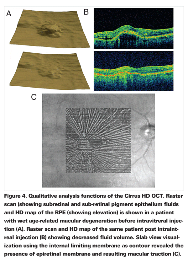
Cirrus HD-OCT (Carl Zeiss Meditec) is a spectral-domain OCT with a speed of 27,000 A-scans/second and a resolution of 5 µm. It utilizes a SLD light source at 840 nm. The design of the Cirrus is different from the other commercial OCT devices since it is not designed around a slit lamp and joystick, but instead employs a mouse. Additionally, the Cirrus has a dedicated iris CCD (charge-coupled device) camera that allows for live monitoring of the pupil during scans and a separate near infrared fundus SLD camera (750 nm) for live fundus visualization. For repeat scans, Cirrus HD-OCT has a "repeat function," whereby with a click of a mouse, the device returns to the previous setup for that particular patient (head and chin rest positions and camera focus) and it activates visit-to-visit registration using retinal vessel tracing. The use of vessel registration is an improvement from older generation devices, as it does not rely merely on the proper patient fixation, thus improving inter-scan reproducibility. The use of registration is also useful for tracking the progression of various ocular diseases. In addition, the Cirrus software provides qualitative HD 3D maps of the ILM and RPE (See Figures 4A & B).
"Slab view" is a unique visualization method that generates OCT enface images from various movable contours (Figure 4 C). This function allows for visualization of pathologies such as epiretinal membranes or macular holes. With the Cirrus HD-OCT the operator can also create customized scan patterns by varying the raster length or scan density and changing the angle of the raster lines.
Copernicus HR
The technologically advanced Copernicus HR (Optopol Technology) delivers the highest scan speed in a commercial OCT system, with an imaging speed of up to 52,000 A-scans/second. This is considerably faster compared to its closest competitor, the Spectralis, with a maximal scan speed at 40,000 A-scans/second. It also has the highest resolution in a commercial device, at 3 µm. The system uses SLD light source centered at 855 nm. The rapid scanning speed decreases scan time and further reduces instances of fixation drift and motion artifacts, while expanding retinal coverage. The improved resolution theoretically allows for the visualization of even higher level of detail. Copernicus HR also has a Doppler analysis module to allow for the examination of the retinal vascular system, and allows for analysis such as flow velocity estimation, and velocity distribution mapping. An anterior segment module is also available for 5 µm resolution imaging of the cornea and anterior segment.
RTVue-100
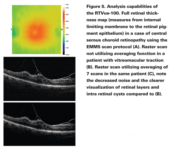
RTVue-100 (Optovue) was the first commercially available spectral-domain OCT device to receive FDA approval. It has a scanning speed of 26,000 A-scans/second and utilizes a low-coherence SLD light source at a near infrared wavelength centered at 820 nm and a bandwidth of 50 nm, achieving a 5 µm axial resolution in tissue. One advantage of the RTVue-100 is its innovative method of image acquisition. The RTVue-100 acquires images in real time, which is particularly useful in patients who are prone to inattention, fixation losses or blinking during scans. Because the software allows the operator to view sets of images taken at different periods of time immediately, it maximizes the opportunity to obtain the highest quality OCT images. Another one of RTVue-100's strengths lies in the wide number of available scan protocols. This diversity provides users in both research and clinical settings multiple ways to view and analyze the retinal layers and optic nerve. For example, EMM5 is the RTVue-100's primary macular mapping scan— it utilizes a grid-like scanning pattern with an outer 6 mm x 6 mm area with 13 horizontal and 13 vertical scans of 0.5 mm interval and an inner 4 mm x 4 mm area with 8 horizontal and vertical scans of 0.5 mm interval. This pattern allows for visualization of even finer details closer to the center of the fovea and creates accurate retinal thickness maps since little interpolation of points is needed. The EMM5 also has the ability to create full retinal, inner and outer retinal thickness maps (Figure 5 A). The wide 6 mm x 6 mm scan area of the protocol provides a high degree of freedom for the operator to reposition the 5 mm-diameter Early Treatment of Diabetes Retinopathy Study-like map in cases of fixation drift. Registration via vessel tracing is also available. Special "high definition" protocols oversample certain points on the OCT scans to decrease speckle (See Figures 5 B & C). The RTVue-100 has an option for an anterior chamber attachment lens, which allows for examination of the cornea and the anterior chamber.
Spectral OCT/SLO
The Spectral OCT (Ophthalmic Technologies) uses a SLD light source centered at 830 to 840 nm to achieve an 8 µm resolution; it has an imaging speed of 27,000 A-scans/second. In addition to OCT, this device also has a confocal scanning ophthalmoscope which uses the same optics as the OCT scanner. The OCT and the confocal SLO generate images that correspond pixel to pixel. Confocal SLO generates the fine surface detail of the fundus, while the OCT C-scan (enface view of a series of sequential OCT scans down the fundus put together) provides the details for the retinal layers. The available registration method of the Spectral OCT/SLO is considerably faster compared to that of other commercial devices since OCT scans are registered to confocal SLO images, which can be generated quickly, instead of to an enface image generated from OCT C-scans. Thus, this system registers scans point-to-point in real time. In addition, OCT/SLO also has an available microperimetry option (which is visual fields over smaller areas of the retina to test patients' ability to perceive lights of various intensities). The data are mapped onto the confocal SLO fundus image and may be also mapped onto the retinal thickness map. In this way, one can assess the relationship between retinal function and structure at particular points in the retina.
Spectralis HRA+OCT
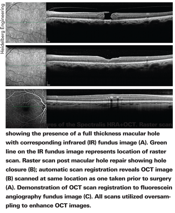
The Spectralis HRA+OCT (Heidelberg Engineering) is one of the most versatile commercially available spectral-domain OCTs. HRA (Heidelberg Retinal Angiography) uses infrared confocal laser scanning for digital acquisition of fluorescein angiography and indocyanine green angiography with 3D resolution. This system allows for up to six different imaging modalities including spectral-domain OCT, fluorescein angiography, indocyanine green angiography, fundus autofluorescence, red free and infrared photography (See Figure 6). The relative high price of the Spectralis due to the various optional factures that may be added onto the core unit (consisting of two imaging modes: SD OCT and infrared photography) presents as a potential disadvantage, but the base units alone are also available. The device has a scanning speed of 40,000 A-scans/second. In addition, it has an axial resolution of 7 µm. The light source used is SLD centered at a wavelength of 870 nm. The Spectralis utilizes a dual-beam scanning system consisting of a confocal SLO reference beam to acquire reference scans for eye movement tracking and guides a second beam to simultaneously acquire OCT images.
The eye-tracking technology recognizes the presence of eye movement then repositions the scan pattern and discards scans with motion artifacts. The Spectralis uses registration by retinal structures to allows for automatic rescan of the retina at the same location as the previous visit, eliminating subjective scan positioning by the operator and thus precise longitudinal tracking of various retinal diseases such as wet age-related macular degeneration is possible. In addition, the Spectralis also over samples specific points on the OCT scans and subsequently compare and combine them to reduce speckle (or random noise) and enhance specific structures.
Factors to Consider
Multiple factors should come into play when considering what is the right OCT device for a particular practice. In clinical settings where there are no dedicated photography departments with skilled operators trained in OCT image acquisition, the ease of image acquisition may be a major concern. For example, Cirrus HD-OCT, with the integration of the mouse and the removal of the joystick may be appropriate. Second, in settings with a high volume of patients seen per day, chair time for the test may become an important consideration and so choosing a device with short image acquisition time may be a good choice. Additionally, sometimes the clinician must weigh the cost of the device against other considerations such as the quality of images obtainable from the device. The spatial dimension of the device might be an important consideration in clinical settings where space is limited. Cirrus HD-OCT is a good example of a compact device, since the monitor and OCT scanners are all bundled into one unit. The type of built-in ancillary devices should also be another factor in choosing an OCT device when different specializations present or financial constraints may become important. Spectralis is a good example of a device in which numerous ancillary devices are available.
Another point of consideration may be based on the methods of OCT scan reporting, where a clinician might decide whether if OCT reports via paper print-outs or direct computer monitor analysis is more effective. In a practice where IMAGEnet is available, Topcon 3D OCT 1000 might be a good choice due to the seamless integration of the two software and the more complete set of information obtainable with computer analysis of scans. Also, the types of qualitative analysis software (such as the "slab view" option on the Cirrus HD-OCT or macular 3D reconstruction on a number of spectral-domain OCT devices) or quantitative analysis software (like volumetric analysis available on the RTVue 100) may be considered based on individual needs and preferences. Lastly, the quality of the segmentation software may be an important consideration in busy practices where manual correction of segmentation lines due to segmentation line breakdown might not be feasible. Having high quality segmentation software is important since it is needed to obtain accurate macular thickness measurements.
Commercial OCT device companies all utilize different segmentation software, and so the rate of segmentation line breakdown also varies across devices; this is especially true in pathological states. Clinicians need to be aware of the differences but also understand that companies regularly update their segmentation software to improve the accuracy of retinal thickness measurements.
Dr. Reichel is director of the Vitreoretinal Service and vice chair,
1. Huang D, Swanson EA, Lin CP, et al. Optical coherence tomography. Science. 1991;254(5035):1178-81.
2. Hee MR, Izatt JA, Swanson EA, et al. Optical coherence tomography of the human retina. Arch Ophthalmol 1995;113:325-32.
3. Schuman, JS, Hee, MR, Arya, AV, et al. Optical coherence tomography: a new tool for glaucoma diagnosis. Curr Opin Ophthalmol 1995; 6(2):89-95.
4. Izatt JA, Hee MR, Swanson EA, et al. Micrometer-scale resolution imaging of the anterior eye in vivo with optical coherence tomography. Arch Ophthalmol 1994;11:1584-9.
5. Wojtkowski, M, Srinivasan, V, Fujimoto, JG, et al. Three-dimensional retinal imaging with high-speed ultrahigh-resolution optical coherence tomography. Ophthalmology 2005; 112:1734-46.
6. Srinivasan, VJ, Adler, DC, Chen, Y, et Al. Ultrahigh-Speed Optical Co-herence Tomography for Three-Dimensional and En Face Imaging of the Retina and Optic Nerve Head. Invest Ophthalmol Vis Sci 2008; 49: 5103-10.
7. Ho, J, Sull, AC, Voung, LN, et al. Assessment of Artifacts and Reproducibility in Pathological Eyes across Spectral and Time Domain Optical Coherence Tomography Devices. Manuscript in submission.
8. Leung, CK, Cheung, CY, Weinreb RN, et al. Comparison of macular thickness measurements between time domain and spectral domain optical coherence tomography. Invest Ophthalmol Vis Sci 2008; 49(11): 4893-7.
9. Forooghian, F, Cukras, C, Meyerle, CB, et al. Evaluation of Time Domain and Spectral Domain Optical Coherence Tomography in the Measurement of Diabetic Macular Edema. Invest Ophthalmol Vis Sci. 2008; 49: 4290-96.