|
Evaluation
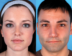
The evaluation of the cosmetic brow patient should include thorough questioning to elicit the patient’s subjective aesthetic complaints, a history of previous facial surgery and any ocular abnormalities. This is followed by a complete head and neck and ophthalmologic exam. Preoperative facial photography should be obtained to assist in performing a comprehensive facial analysis to include an assessment of brow position. The brow should lie roughly 1 cm above the medial canthus along an imaginary line perpendicular to the nasal ala, and end laterally at an oblique line extending from the alar ridge through the lateral canthus. Although debated, the highest point of the brow should generally be near the lateral limbus, and the arc is more pronounced in the female (See Figure 1). The female brow should lay 0.5 to 1 cm above the supraorbital ridge, while the male brow should be located at the level of the supraorbital ridge.
Anatomy & Surgical Technique
Browlifting addresses brow ptosis primarily, but some techniques may impact forehead and periorbital rhytids. Structures at risk during brow and forehead lifts include the neurovascular bundles emanating from the supraorbital region, as well as the temporal (or frontal) branch of the facial nerve. It is imperative to understand the neurovascular anatomy of the forehead to avoid damage to these structures intraoperatively. The layers of the scalp include the skin, connective tissue, galea aponeurotica, loose areolar tissue and the periosteal layer. The superficial temporal artery supplies the lateral forehead, while the medial forehead receives its vascular supply from the supraorbital and supratrochlear arteries, both branches of the ophthalmic artery (which itself arises from the internal carotid artery). The medial forehead receives its sensory supply via the supraorbital and supratrochlear nerves from the V1 branch of the trigeminal nerve, while the lacrimal (V1), zygomaticofacial (V2) and the auriculotemporal (V3) address the lateral forehead sensation.
The temporal branch of the facial nerve courses superomedially through the area between the brow and hairline within the temporoparietal fascia (TPF). Anatomic studies revealed that the sentinel vein, a structure encountered in the sub-temporoparietal fascial plane in the temple, is a reliable predictor of the nerve’s course, and runs inferior to the nerve within 2 mm2. Forehead and periorbital rhytids are caused by contraction of the upper facial musculature, including the corrugator, frontalis and procerus muscles. The corrugator muscles are small, fan-shaped muscles that underlie the eyebrows and cause vertical glabellar wrinkling. The frontalis muscle is a broad, bi-lobed muscle that raises the eyebrows. The procerus extends from the dorsal nose to the lower forehead, and is responsible for upper nose wrinkling.
Here are the different surgical options for treating the brow and forehead, in order of popularity in our practice.
Endoscopic Browlift
In our opinion, the endoscopic browlift is the procedure of choice for rejuvenating the brow. The advantages of the endoscopic browlift include its minimally invasive nature and the absence of long incisions; the inherent ability to provide a natural result and avoid overly-elevated brows; the ability to directly visualize the procerus and corrugator muscles for resection; and the low incidence of scalp paresthesia. Disadvantages include the need for anesthesia; costs associated with fixation; and a posterior hairline shift.
|
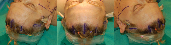
A full discussion of surgical detail is not possible in this review, but we will provide a brief review of our technique. We most often combine the endoscopic browlift with the temporal browlift described hereafter. Five scalp incisions are made posterior to the hairline; one midline, two in the paramedian position over the brow arch, and two that are parallel to the hairline in the temple (See Figure 2).
Periosteal elevators are used to dissect in the subperiosteal plane to the supraorbital ridge, avoiding the supraorbital and supratrochlear nerves. The temporal browlift is completed as described below in the sub-TPF plane. The temporal dissection is connected to the frontal dissection by dividing the conjoint fascia, or temporal line. This is done through the temporal pocket in a lateral to medial fashion, which protects the facial nerve. Around the glabellar and superior orbital rim areas the periosteum is divided transversely with reverse elevators or endoscopic scissors. It should be noted that frontal paresis occurs more commonly on the left because on this side the endoscope is positioned directly under the frontal branch.
|
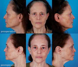
Once the tissue is released from periosteum, it is redraped and fixed posteriorly after adequate flap advancement. There are several flap fixation techniques, generally using screws or other bioabsorbable soft tissue fixation devices. We prefer fixation screws, which are removed 10 to 14 days postoperatively. Periosteal reattachment is stable within six to eight weeks (See Figure 3).
Temporal Browlift
The temporal browlift can be performed in conjunction with the endoscopic browlift or as a separate procedure to address lateral brow and periorbital rhytids. There are many different ways to perform a temporal browlift, from endoscopic (most commonly in conjunction with a full browlift) to subcutaneous (most commonly an office procedure under local anesthesia). We will describe the endoscopic approach. An incision is made 8 to 10 mm posterior to hairline over the temporal suture line. Dissection is carried down to the superficial layer of the deep temporal fascia, under the temporoparietal fascia. Using sharp elevators, care is taken to stay in this plane as dissection proceeds towards the lateral orbital rim. The sentinel vein should be identified and preserved if possible. If it is necessary to ligate the sentinel vein, this should be done as close to the temporalis muscle as possible to avoid injury to the facial nerve. Ligation may increase postoperative edema.
|
Direct Browlift
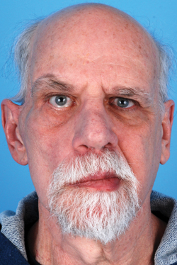
The direct browlift is most useful in the comprehensive management of facial paralysis and in male patients with prominent rhytids. It is rarely used for cosmetic patients in our practice. Separate incisions are made over each brow; the inferior edge of each incision is placed within the superior most aspect of the eyebrows, and carried to the lateral aspect of the eyebrow and extended horizontally in a gentle arc. It should not extend more medially than the medial aspect of the eyebrow as this may result in glabellar scarring. The superior incision should be placed a maximum of 10 to 12 mm above the inferior incision, with its highest point at the lateral limbus. The superior incision determines the new brow positioning, and the degree of femininity or masculinity of the rejuvenated face depends on the angle of the eyebrow at the lateral limbus. This is a powerful technique and care the surgeon should err on the conservative side to avoid an over-elevated brow (See Figure 5). The advantages of a direct browlift include its short operative duration, the ability to perform under local anesthesia, and its precise control over the design of brow contour and shape. It does not change the appearance of forehead or glabellar rhytids, and does leave a visible eyebrow scar.
Midforehead Lift
This technique utilizes deep forehead furrows via a transverse midforehead incision. As with the direct lift, this technique is rarely indicated in the female cosmetic patient. It is indicated almost exclusively for men and for patients that may benefit from forward advancement of the hairline.
|
Coronal Forehead Lift
The coronal lift is useful for situations in which endoscopic procedures have failed or are not indicated or for patients who do not require a pretrichial incision. In our practice, this situation is exceedingly rare. The advantages of the coronal lift include the ability to address the corrugator, procerus and frontalis muscles to theoretically eliminate dynamic rhytids; the wide surgical exposure for teaching purposes; and the possibility to powerfully elevate the brow. Two major disadvantages include hairline elevation and paresthesia posterior to the incision.
An incision is placed posterior to the hairline in an arcuate design towards the helical root. Dissection is carried out in the subgaleal plane to the superior orbital rim, with attention to the course of the superficial temporal branch of the facial nerve. The corrugator muscles are dissected out and removed with sharp scissor dissection, avoiding the supratrochlear nerve. The procerus muscle can be similarly addressed if desired. Replacing the flap is carried out with relaxing incisions and cardinal sutures placed in areas requiring the greatest amount of lift. After satisfactory replacement of the brow, redundant areas of scalp skin are excised. Galeal sutures are placed in several areas along the incision, with emphasis along the dome of the head to avoid postoperative scar spread.
Pretrichial Browlift
The pretrichial browlift is indicated in women with high hairlines or for long vertical forehead heights. It is a modification of the coronal lift, and does not lift the anterior hairline further.
|
Browpexy
A browpexy may be used in combination with the upper blepharoplasty and may negate the need for a forehead procedure. It cannot address forehead rhytids or other aesthetic concerns of the upper third of the face. It is performed through an upper blepharoplasty incision after identification of the supraorbital vessels and nerves surrounding the supraorbital notch. The blepharoplasty incision is extended superiorly to 1.5 cm above the superior and lateral orbital rim deep to the orbicularis oculi muscle. Blepharoplasty is carried out first, then browpexy is carried out to elevate and suspend the brow. Sutures are placed at the infrabrow hairs and passed in the periosteal plane to 1 cm above the supraorbital ridge. Tying down these sutures will elevate the eyebrow, and therefore placement and tension of the suture guides repositioning of the brow.
Complications
Main complications of the browlift procedures include bleeding, injury to sensory or motor nerves, lagophthalmos and alopecia. Bleeding can occur from the superficial temporal artery and zygomaticotemporal artery, as well as the supratrochlear and supraorbital arteries. Given the close proximity to nerves throughout the lateral forehead, cautery with the bipolar is recommended. The development of postoperative hematoma could compromise flap vascularity and survival, and is an indication for urgent exploration and cautery. Stretching of the supratrochlear and/or supraorbital nerve can result in temporary hypesthesia over the supraorbital rim. In the direct browlift, permanent hypesthesia generally results surrounding the incision. The browpexy is also associated with hypesthesia, generally of several months’ duration, over the lateral eyebrow margin.
|
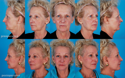
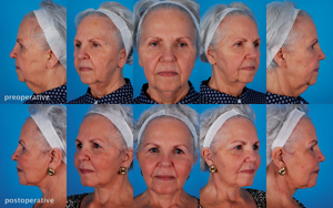
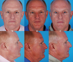
Although the surgical browlift seems less popular in favor of volume replacement techniques recently, it remains a powerful rejuvenative technique in the plastic surgeon’s arsenal. It can beautifully complement other facial cosmetic procedures (See Figure 7). There are several procedures to address the aesthetic aspects of the upper third of the face. Each should be tailored to the specific needs of the patient undergoing the procedure. Choice of technique will depend on the position of the patient’s hairline, the amount of lift that is necessary, and the need to concurrently address forehead rhytids or brow asymmetries. Only browlifts were discussed in this review, however, a thorough consideration of all aspects of the individual patient and the aging forehead, brow and eyelids should be undertaken in order to optimize facial rejuvenation. REVIEW
Dr. Heffelfinger is the director of the Division of Facial Plastic and Reconstructive Surgery and co-director of the Herbert Kean Center for Facial Aesthetics at Thomas Jefferson University Hospital. He also the director of Head and Neck Microvascular Surgery at Jefferson. Contact him at 925 Chestnut St. 6th fl. Philadelphia, PA 19107. E-mail: ryan.heffelfinger@jefferson.edu. Dr. D’Souza is a third-year resident in Jefferson’s otolaryngology residency program. Contact her at jillndsouza@gmail.com.
1. Codner MA, Kikkawa DO, Korn BS, Pacella SJ. Blepharoplasty and Brow Lift Plast Reconstr Surg 2010;126(1):1-17.
2. Sabini P, Wayne I, Quatela V. (2003) Anatomical Guides to Precisely Localize the Frontal Branch of the Facial Nerve. Arch Facial Plast Surg 2003;5(2):150-152.
3. Adamson P, Dahiya R. The Aging Forehead. In: Bailey B, Johnson J. eds. Head and Neck Surgery—Otolaryngology. Philadelphia: Lippincott Williams & Wilkins 4th Ed, 2006;2663-2683.
4. Paul M. The Evolution of the Brow Lift in Aesthetic Plastic Surgery. Plast Reconstr Surg 2001;108:1409-1424.
5. Graham DW, Heller J, Kurkjian TJ, Schaub TS, Rohrich RJ. Brow Lift in Facial Rejuvenation: A Systematic Literature Review of Open versus Endoscopic Techniques. Plast Reconstr Surg 2011;128(4):335-341.
6. Vinas JC, Caviglia C, Cortinas JL. Forehead Rhytidoplasty and Brow Lifting. Plast Reconstr Surg 1976;57:445.
7. Knize DM (2009) Anatomic Concepts for Brow Lift Procedures. Plast Reconstr Surg 124(6):2118-2126.
8. Agarwal CA, Mendenhall SD 3rd, Foreman KB, Owsley JQ. The Course of the Frontal Branch of the Facial Nerve in Relation to Fascial Planes: An Anatomic Study. Plast Reconstr Surg 2010;125(2):532-7.
9. McKinney P, Mossie RD, Zukowski ML. (1991) Criteria for the Forehead Lift. Aesthetic Plast Surg 15:141-147.
10. Vasconez IO, de la Torre JI. (2002) Fine Tuning the Endoscopic Brow Lift. Aesthet Surg J 2002;22:69-71.
11. Nassif PS. Kokoska MS, Homan S. (1998) Comparison of subperiosteal vs subgaleal elevation techniques used in forehead lifts. Arch Otolaryngol Head Neck Surg 1998;124(11):1209-1215.
12. Mackay GJ, Nahai F. (1995) The Endoscopic Forehead Lift. Operative techniques in plastic and reconstructive surgery. Plast Reconstr Surg 1995; 2(2):137-144.
13. Matarasso A, Hutchinson O. Evaluating Rejuvenation of the Forehead and Brow—an Algorithm for Selecting the Appropriate Technique. Plast Reconstr Surg 2000;106: 687-689.
14. Elkwood A, Matarasso A, Rankin M. (2001) National Plastic Surgery Survey: Brow Lifting Techniques and Complications. Plast Reconstr Surg 2001;108:2143-2150.
15. Byun S, Mukovozov I, Farrokhyar F, Thoma A. Complications in Brow Lift Techniques: A Systematic Review. Plast Reconstr Surg 2012;130:90.
16. Swanson E. Objective Assessment of Change in Apparent Age after Facial Rejuvenation Surgery. J Plast Reconstr Aesthet Surg 2011; 64(9):1124-1131.
17. Angelos PC, Stallworth CL, Wang TD. 2011 Forehead Lifting: State of the Art. Facial Plast Surg 2011;27(1):50-57.


