From its first appearance, frequency-doubling technology has shown the ability to reveal early defects in glaucomatous visual fields. While the first FDT instruments confirmed this technology's potential, they never achieved sufficiently widespread clinical use to make them a serious challenger to standard automated perimetry as the gold standard for visual fields. But the Humphrey Matrix Perimeter with Welch Allyn frequency-doubling technology from Carl Zeiss Meditec has made far greater inroads into clinical practice.
The Matrix offers three main testing algorithms for generating visual fields. One is a screening algorithm similar to SITA-Fast where parts of the visual field are not retested, allowing you to cover the visual field quickly. It also can perform central 10-degree testing, which can be used to follow a patient with advanced glaucoma, and a more mainstream 24-2 algorithm that's comparable to 24-2 SITA SWAP.
Using FDT in Practice
Christopher A. Girkin, MD, a professor of ophthalmology and director of the glaucoma service in the Department of Ophthalmology at
"In clinical practice, I use the Matrix in patients who have normal visual fields on standard perimetry but optic nerves that are suspicious for early glaucoma," he explains.
"I wouldn't use the Matrix as a basis for treatment without confirmation of its findings; our studies of both types of perimetry have found that false positives occur about 10 percent of the time. If I see a defect on a Matrix field that makes sense—by which I mean that it fits with what I see when I examine the optic nerve—that would make me suspicious that the patient may actually have early glaucoma. For example, let's say there's a little thinning of the inferior temporal rim with some corresponding beta zone atrophy. It looks a little suspicious, but it's not overtly abnormal. If I then see a superior arcuate defect on FDT with a full standard visual field, I think that's informative. The key is to be sure that Matrix findings correspond to and corroborate other information."
George Shafranov, MD, clinical associate professor of ophthalmology at the Yale University School of Medicine, and in private practice in
"It may turn out that many of these patients showing defects on Matrix will develop glaucoma in the future," he continues. "Matrix may be so sensitive that no other commercially available technology is able to pick up these changes. An analogy might be the Heidelberg Retina Tomograph in the early 1990s. In '94 and '95, some of the patients who were followed on the HRT showed structural progression, but the visual fields and photographs of the time didn't confirm this. As a result, many glaucoma specialists were skeptical about whether the HRT was truly demonstrating early glaucoma progression.
But five or 10 years later, many of the patients picked up by HRT were confirmed on other tests, so the HRT gained widespread acceptance.
"It's possible that the situation with the Humphrey Matrix is similar," he says. "We don't know yet."
In terms of how Matrix data can be used to monitor progression, Dr. Shafranov notes that the lack of a database results in some limitations. "You can sequentially analyze the results of Matrix visual fields," he says. "You can look at individual dots to see if a defect has become deeper or larger. But the significance of that progression is hard to judge because there's no variability algorithm included in the analysis of those changes."
FDT vs. Standard Perimetry
"I don't think it's clear whether the Matrix in its current form is more sensitive than standard perimetry in terms of detecting early disease," says Dr. Girkin. "If you look at most of the clinical studies that have been done using patients who are defined by the condition of the optic nerve, standard perimetry and FDT are roughly equal in sensitivity—although there's some emerging evidence that FDT may be a little bit better at detecting early disease. However, the Matrix appears to be detecting a different, incompletely overlapping subset of early glaucoma patients than those detected with standard perimetry. Several clinical studies are in agreement about that, although the reasons for the difference are not yet clear."
Dr. Girkin notes that one potential disadvantage the Matrix has relative to standard perimetry is larger intervals and fewer steps between threshold changes for monitoring early disease. "It's like having two rulers that are 12 inches long, but one has only 10 marks for measuring," he explains. "Developing a thresholding algorithm that's more sensitive to measuring early disease would be an improvement. I do think the Matrix is useful in this situation, but I don't necessarily think it's more sensitive than standard perimetry, at least in its current form.
"The primary advantage Matrix has over standard perimetry is that its threshold variability is less affected by disease severity," he continues. "In fact, the current design may be better at evaluating moderate to more advanced disease for progression. This may relate to the size of the stimulus; a larger stimulus samples more ganglion cells in the retina and may be more reproducible. This results in a more robust, less noisy test as glaucomatous scotomas worsen, as data in one recent paper suggested.1 You don't get wider variability in damaged areas, as seen with standard visual fields. Of course, this potential FDT advantage needs to be proven with longitudinal data."
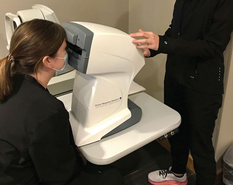
Administering the Test
Dr. Girkin notes that in terms of day-to-day use, the Matrix has pros and cons. "The Matrix is a smaller machine, less cumbersome, easier to use and more resistant to spectacle blur," he points out. "I think most patients tend to prefer the FDT. The downside is that the Matrix is less versatile. The Humphrey Field Analyzer can do a lot, including standard perimetry and SWAP, and you can customize it to do a variety of different tests. The Matrix can also perform a variety of tests, but you're a little more constrained."
Despite its limitations, Dr. Shafranov says there are good reasons for a practice to own a Matrix. "The Matrix visual field test is very easy to administer," he notes. "The instrument itself takes up significantly less space than the Humphrey visual field analyzer, and the sensitivity of Matrix is pretty much as high as SWAP. It's a good screening tool, and patients seem to prefer it to the regular Humphrey analyzer. For physicians who want to follow patients in their own offices rather than providing their opinion on a visual field progression to somebody else, Matrix is an excellent option."
Dr. Shafranov says patients appear to prefer it over standard visual field testing because it's less stressful and more comfortable. "It's easier to see the stimuli because they flicker," he notes. "In contrast, standard perimetry uses a very faint light that only stays on the screen for 0.2 seconds; patients aren't always sure whether they saw it or not.
They're more certain that they pushed the button correctly during the Matrix test. The test also takes somewhat less time—three to five minutes, depending on the complexity of the field. Standard perimetry takes four to seven minutes. And physically, Matrix is easier on the patient. Patients just look inside a tube instead of trying to position their head in a chin rest, which can stress the neck and back."
Dr. Shafranov notes that the Humphrey Matrix compares better to SAP than the original FDT instrument did. "The Matrix allows resolution identical to what standard automated perimetry gives us," he says. "That was the main advantage standard automated perimetry had over the original FDT (from Welch Allyn); it tested more spots in the visual field. Matrix allows higher resolution of visual field defects. In fact, Matrix appears to be more sensitive than automated perimetry and the SWAP visual field test that's administered on the Humphrey Field Visual Analyzer II. Also, it tests slightly different parts and functions of the ganglion cells than the original FDT instrument.
"In any case, comparative studies of SAP and the Matrix have not found either one to be significantly more sensitive. That gives more weight to practical considerations, where the Matrix has significant advantages in terms of administering the test. So I don't see any reason not to use the Matrix, except that we don't have the standardized databases that help with long-term analysis."
A Worthwhile Practice Investment?
Dr. Shafranov believes the Matrix is worth adding to a practice, especially if the doctor is not a glaucoma specialist. "In many respects, Matrix interpretation is easier than SAP interpretation," he notes. "Defects are more defined and localized; diffused change is not seen as often as in SAP. However, I don't know if information from Matrix can be easily transferred to other offices and interpreted the same way at a different location, the way SAP can.
"I see the Matrix as a second device to purchase so you can come up with your own opinion," he continues. "You can perform both tests and compare the results. But until national studies are conducted in multiple centers with independent readers to interpret the results of the two tests with blind data, I don't think Matrix will gain as wide acceptance as SAP. I'm not aware of any such studies at the moment. But once such data do exist, my feeling is that Matrix might replace SAP."
"I'd be very much inclined to switch to the Matrix," he adds. "It's a much easier test. It's the size of a small printer with a very small footprint. You don't need a special room for it; you can put it at the edge of a table. And from a practical standpoint, if I had to choose a visual field to perform on myself, I'd choose the Matrix. I've taken both, and it's an easier and more comfortable test."
Asked whether a Humphrey Matrix is a worthwhile investment, Dr. Girkin says it depends. "If your practice sees a lot of glaucoma and you want another test to evaluate suspects, I think it's worthwhile," he says. "I use it. But I don't think it's the end-all, be-all of glaucoma management, and I don't think it's necessary to manage glaucoma patients appropriately. It's certainly not the gold standard, though it conceivably could be at some point in the future. It has advantages and disadvantages, so it's an individual practice decision. It would be greatly enhanced by modifying the software to allow for increased threshold steps in early loss."
What Lies Ahead?
Dr. Girkin notes that several ongoing large-scale clinical trials are accumulating Matrix data as part of their protocols. "We're involved in one—the African Descent and Glaucoma Evaluation Study (ADAGES)—that's following a large cohort of African-American patients and patients of European ancestry to develop progression algorithms," he says. "This study is being conducted with the
Dr. Shafranov believes that the Matrix's version of frequency-doubling testing still has a few hurdles to overcome. "Most of the national prospective glaucoma studies have used standard automated perimetry," he notes. "For that reason, it's hard to see Matrix replacing standard perimetry right now. But health-care costs will be an issue in the future, and the Matrix exam takes significantly less time, potentially leading to greater efficiency. It also creates fewer opportunities for human error and allows easier training for the technician. Head position in standard automated perimetry can cause more artifacts than with the Matrix. And, the Matrix stresses patients less; since it's a psychophysical test, patients' overall awareness of their answers makes the Matrix exam easier. Given those factors, Matrix use should continue to increase, especially once we have Matrix databases similar to those that now exist for standard auto perimetry and SWAP analysis."
Dr. Shafranov says that when the Matrix was first introduced he thought it would be an ultimate answer to visual field testing. "I still believe it's more sensitive than standard automated perimetry," he says, "but we practice in an age in which standards of care dominate over any as-yet-unproven new device. So far, no large, prospective study has conclusively demonstrated that Matrix is reliably more sensitive than other tests. At least one paper has found that SAP and the Matrix may identify different groups of eyes as abnormal,2 so perhaps using both machines is the most appropriate option right now. Given the broad acceptance of standard automated perimetry, it doesn't seem reasonable to stop using it."
"It's possible that the Matrix could eventually replace standard perimetry," agrees Dr. Girkin. "Several factors will play into that. One is the issue about the smaller number of threshold steps per change, which could turn out to be a disadvantage for the Matrix. On the other hand, it appears to be more robust and less noisy as a test for advanced disease, which may work in its favor. Another consideration is that standard perimetry is so heavily used; there's a lot of existing data on it. There are also new devices coming out, including technology such as SITA SWAP and
Dr. Girkin receives research support from Carl Zeiss Meditec and Heidelberg Engineering. Dr. Shafranov has no financial interest in any of the products discussed.
1. Wall M, Woodward KR, Doyle CK, Artes PH. Repeatability of Automated Perimetry: A Comparison between Standard Automated Perimetry with Stimulus Size III and V, Matrix, and Motion Perimetry. Invest Ophthalmol Vis Sci 2009;50:974-979.
2. Sakata LM, Deleon-Ortega J, Arthur SN, Monheit BE, Girkin CA. Detecting visual function abnormalities using the Swedish interactive threshold algorithm and matrix perimetry in eyes with glaucomatous appearance of the optic disc. Arch Ophthalmol 2007;125:3:340-5.