Presentation
A 21-year-old African-American female presented to the Wills Eye Emergency Room complaining of sudden, painless, bilateral decreased vision, left greater than right. She stated her symptoms began three days prior while on vacation, at which time she reported decreased water intake. She denied any pain with eye movement or other systemic symptoms.
Medical History
| ||||
Examination
Ocular examination revealed visual acuity of 20/25 in the right eye and 20/200 in the left eye. Pupils were equal and reactive, and there was no afferent pupillary defect. Ocular motility in both eyes was full. On confrontation visual fields, there was a small defect in the superior field on the right and the temporal field on the left. Applanation tonometry measured an intraocular pressure of 14 mHg on the right and 12 mmHg on the left. The patient read seven out of eight color plates on the right and eight out of eight color plates on the left, both with some difficulty.
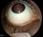
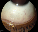
Slit-exam examination revealed unremarkable external and adnexal structures. The scleral and conjunctival exam exhibited a positive comma sign in the inferior fornix of both eyes (See Figures 1a & b). The remainder of the anterior segment exam was otherwise normal.
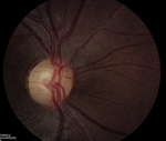
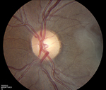
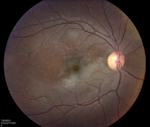
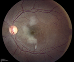
Posteriorly, the vitreous was clear in both eyes. Segmented disc capillaries were present on both optic nerves (See Figures 2a & b). There was an area of retinal whitening in the posterior pole of each eye (See Figures 3a & b). No thrombus was visualized, and the periphery revealed no salmon patch hemorrhages or neovascularization.
| ||||||||||
What is your differential diagnosis? What further workup would you pursue?


