Presentation
A 54-year-old Caucasian male presented to the Neuro-Ophthalmology Service at Wills Eye for peripheral visual disturbances in his right eye. The patient noted painless blurry vision in his superior visual field that started suddenly, approximately 10 days prior to his visit. During that time, the patient believed that the blurry vision had progressively worsened. The patient denied headaches, jaw claudication, fever and weight loss. Review of systems was negative.
|
The patient’s medical history was positive only for hyperlipidemia for which he takes atorvastatin. His ocular history was negative.
Examination
The patient’s visual acuity without correction was 20/25 in the affected eye and 20/20 in the left eye. A relative afferent pupillary defect was noted on the right. Extraocular motility was intact in both eyes. Color plates were 5.5/8 in the right eye and 8/8 briskly in the left eye. Slit-lamp examination was unremarkable. Humphrey visual fields revealed a superior altitudinal defect in the right eye and a normal field in the left eye; however, the test was complicated by a high number of fixation losses and increased false negative responses.
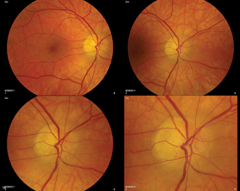
The right optic nerve revealed edema inferiorly (See Figure 1). No disc hemorrhages were noted. The left optic nerve was normal with a cup-to-disc ratio of approximately 0.4. The remainder of the posterior exam was unremarkable in both eyes.
What is your differential diagnosis? What further workup would you pursue?