A 77-year-old Caucasian man presented to the Wills Eye Emergency Room because of blurred vision in his right eye for the previous four hours. He described his vision loss as a “white cloud” covering the entire visual field in his right eye. This was the fourth episode of similar symptoms for him during the past year, with vision loss each time lasting from three to 48 hours. He had not noted any flashes or floaters, headache, temporal tenderness, pain on chewing or muscle weakness.
Medical History
The patient had polymyalgia rheumatica, for which he took 5 mg of oral prednisone daily for maintenance therapy. Cataract extraction in both eyes was performed four years earlier with implantation of a posterior chamber intraocular lens in his right eye and an anterior chamber intraocular lens in his left eye.
The first two of the prior episodes of “white out” happened one month apart approximately one year prior. The first, which lasted four hours, occurred when he went outside into bright sunlight. Shortly after that, he saw his ophthalmologist, who did not detect any pathology. The second episode began while he was inside a movie theater, and lasted approximately 48 hours. He then came to the Wills Eye Emergency Room after resolution of symptoms. Examination at that time was unremarkable. After phone consultation with neuro-ophthalmology, labs were ordered to rule out Giant Cell Arteritis; erythrocyte sedimentation rate was 19; C-reactive protein was elevated at 3.4; and platelets were 190. Because of left upper extremity weakness and headache, he was referred to the main ER for stroke evaluation, which was negative.
The following week his neurologist diagnosed him with GCA and instructed him to increase his oral prednisone from 5 mg/d to 40 mg/d. He was subsequently referred to multiple specialists and, in addition to being told to stop his increased prednisone dose, he had carotid dopplers, a head/neck MRI/MRA, and echocardiography looking for causes of amaurosis fugax, vertebrobasilar insufficiency, or cardiac arrhythmia. All studies were negative. He was asymptomatic for about 10 months, at which point he had a third episode of cloudy vision in his right eye lasting three hours. The following month he experienced his fourth attack and, for the first time, presented for evaluation while symptomatic.
Examination
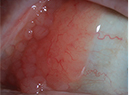
Best-corrected visual acuity was hand motion only in the right eye and 20/50 in the left. Extraocular motility was full. The pupils were irregular (surgical changes after cataract surgery). No relative afferent pupillary defect was present. Intraocular pressure was 34 mmHg in the right eye and 12 mmHg in the left. The anterior chamber of the right eye was filled with 4+ pigment and 4+ red blood cells. Pseudophacodonesis was noted on eye movement. There was a nasal iris transillumination defect in both eyes. The optic nerves and retina appeared normal. Two hours later his right anterior chamber had cleared to 2+ and his acuity improved to 20/70.
What is your differential diagnosis? What further workup would you pursue?