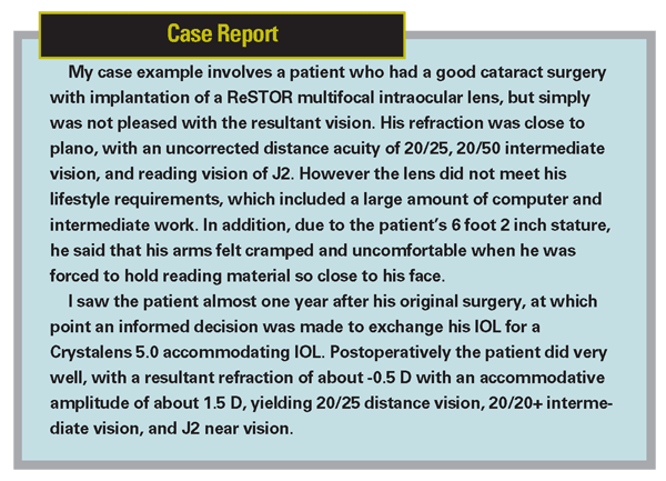
With patients having increasingly high expectations for their vision and spectacle independence, choosing the right intraocular lens for the right patient can be a challenge. No matter how thorough the patient education and how precise the lens calculations, at times the IOL can be mismatched to the patient. In this situation, an IOL exchange may be the best option to deliver satisfaction and good vision to the patient.
Patient Expectations
In the majority of potential IOL exchange candidates, the original lens was likely an appropriate choice but perhaps didn't meet the patient's expectations. Make sure that the patient does not have unrealistic expectations, a goal of 'perfect' vision, or a mindset of finding the fountain of youth. Any additional surgical procedure such as an IOL exchange has higher risks than the original surgery: additional incisions in the eye which can affect astigmatism and healing, further potential for corneal endothelial cell loss, and a repeat exposure to the risks of endophthalmitis and retinal complications. The most important risks that IOL exchange patients face are rupture of the capsular bag and development of cystoid macular edema. Patients need to be patient with the postop recovery, which can be prolonged when compared to routine cataract surgery, and they need to understand that if their capsular bag breaks during surgery, they may receive a different IOL than originally planned.
Preoperative Evaluation
If the first IOL was compatible with the patient's expectations but just resulted in a refractive error, then a simpler procedure, such as a piggy-back IOL or an excimer-based corneal ablation, may be the better choice. If, however, the patient has received an IOL design that he cannot not tolerate, such as a multifocal IOL or a blue-blocker IOL, then an IOL exchange is the more suitable procedure.
IOL calculations are critical and every attempt to get the records of the first surgery should be made, as knowing the power of the original IOL and the resultant postop refraction will allow you to more precisely select your new IOL. If the patient has significant cylinder, try to adjust your incisions or perform additional relaxing incisions to reduce the amount of astigmatism.
At the slit-lamp, note the size and shape of the capsulorhexis, the type of IOL used, and the position of the incisions. Hydrophobic acrylic IOLs tend to be more adherent to the capsular bag than IOLs made from other materials, and as such, they can be more difficult to dissect free.

Medications
Preoperative treatment with non-steroidal anti-inflammatory drugs (NSAIDs) is important for four reasons: 1) prevention of intra-operative miosis; 2) analgesia and patient comfort; 3) reduction in the postop inflammatory response; and most importantly, 4) prevention of postop cystoid macular edema. Because of the additional trauma induced during the second surgery, use of an NSAID can play an important role in preventing postop CME, which would otherwise significantly limit vision. The important considerations for selection of an NSAID include potency, penetration, clinical efficacy and patient compliance. I like to have the patients use the NSAID for six full weeks, so having a drop that is dosed just once or twice a day is more advantageous, particularly when it is very potent and has the ability to penetrate long, myopic eyes.
Surgical Technique
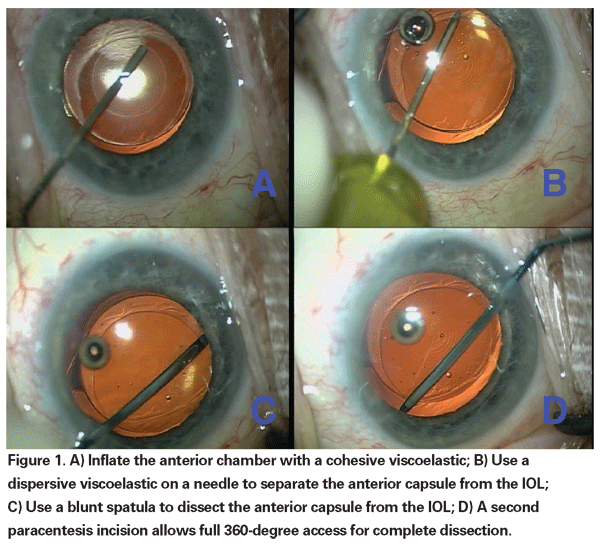
I tend to perform these cases under topical anesthesia with the addition of intrcameral preservative-free lidocaine 1%, as well as intravenous systemic sedation by an anesthesiologist. Fill the anterior chamber with a cohesive viscoelastic to keep it formed during the surgery. Next, use a more dispersive viscoelastic on a 25-ga. or 27-ga. needle instead of a blunt canula. The needle can be placed bevel down and under the edge of the anterior capsular rim. Once it is placed under the capsulorhexis edge, inject to perform viscodissection. Next, use a blunt spatula to fully dissect the anterior capsule free from the IOL. Making a second paracentesis opposite from the first one is helpful, allowing a full 360 degrees of access.
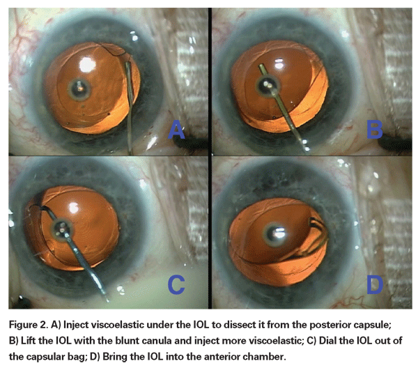
By injecting viscoelastic under the IOL optic, the capsular bag can be safely dissected and separated from the acrylic lens surface. Once there is some protection of the posterior capsule, the blunt viscoelastic can be used to gently lift the IOL. The chopper or second instrument can then be used to dial the IOL out of the capsular bag and into the anterior chamber.
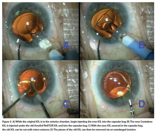
Once the original IOL is in the anterior chamber, bring it up towards the corneal endothelium. The previously injected viscoelastic will be a good barrier to protect the corneal endothelium. At this point, some surgeons like to remove the original IOL before placing the new IOL. In my hands, I have found this to be associated with a higher risk of inadvertently damaging the delicate posterior capsule.
I prefer to inject the new IOL into the capsular bag first, then once it is in position, I can remove the old IOL. The new IOL acts as a barrier to protect the posterior capsule from the sharp tips of my instruments. I prefer to use the MST micro-forceps and micro-scissors to cut the IOL and remove the pieces through the original incision. At this point, the viscoelastic can be removed and the incisions can be secured.
Postoperative Management
The prolonged surgical time and intraocular manipulation can cause more inflammation and make for a longer postop recovery. To help with the inflammation and to speed visual improvement, I like to prescribe a strong steroid in addition to an NSAID. Prednisolone acetate 1% is prescribed four times a day for threeweeks, and Xibrom 0.09% (bromfenac) is prescribed b.i.d. for six weeks. This ensures that the inflammation is fully resolved, the postop discomfort is eliminated, and the risk of cystoid macular edema is reduced.
Because of the extra incisions, opening prior incisions, and the re-operation, there may be a higher risk of incision leakage, and therefore infection, after surgery. Use of a fourth-generation fluoroquinolone is highly recommended both before and after surgery. My goal for the antibiotic is a fast kill time and a full spectrum of microbial coverage, and therefore I typically select Zymar (gatifloxacin).
During the post-operative period a repeat dilated fundus examination is indicated in order to search for possible retinal breaks or weakness that may have been created during surgery as well as to monitor the macula for signs of edema.
An IOL exchange surgery can be challenging due to the increased risks and higher degree of technical intricacy, but it can provide a remedy for patients when the original IOL does not meet their expectations. Educate and inform the patient about the nature and details of the surgery, proceed with care and caution, and choose a new IOL that more closely meets the patient's needs.
Video of this surgery available at:
http://www.youtube.com/results?search_query=%22uday+dev gan%22&search=.
Dr. Devgan is in private practice at the Maloney Vision Institute,
He is a consultant for Allergan, AMO, Bausch & Lomb, Eyeonics, Ista Pharmaceuticals and Staar Surgical, and he has received award funds and travel support from Alcon.


