Although we live in a world of high-tech wonders, sometimes a low-tech alternative can be a powerful tool. Here are two recently developed simple tests that help measure functionality in patients with low vision.
Testing Rudimentary Vision
When a patient’s visual acuity is very poor, most eye-care practitioners use “count fingers” or “hand motion” to categorize it. Recently, a simple new system has made it possible to measure very low vision with much greater precision in two to three minutes using a series of handheld cards. The system was developed by Ian L. Bailey, OD, MS, FAAO, and colleagues. Dr. Bailey is a clinician and researcher at the University of California, Berkeley, where he is also professor of optometry and vision science.
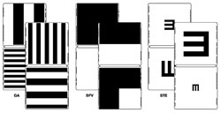
The Berkeley Rudimentary Vision Test adds 13 increments between the lowest acuity level measureable with a standard eye chart and “light perception only.” The test consists of three pairs of hinged cards, 25 cm2, held in front of the patient at prescribed distances. The cards display four single tumbling E optotypes (145 mm, 92 mm, 58 mm and 36 mm); four grating acuity targets (290 mm, 184 mm, 116 mm and 72 mm); a white field projection test; and a black/white discrimination test. Angular size is increased by using short viewing distances.
Dr. Bailey notes that count fingers and hand motion are non-standardized. “People who use count fingers frequently don’t specify the distance, the background or whether the hand is moving or not,” he points out. “Also, its not encouraging for the patient, because the clinician has given up using measurement in the usual way.” Dr. Bailey adds that increased precision matters. “If the patient’s undergoing treatment, you’d like to know whether it’s having an effect or not,” he says. “Count fingers and hand motion can’t identify anything other than very large changes.”
Dr. Bailey says two experiences inspired him to create the new system. First, the World Blind Cricket Council asked for help classifying the severity of visual impairment of their athletes. Shortly thereafter, he attended a think-tank meeting at which scientists working with the latest bionic retinal implants were judging the increase in vision produced by the implants in terms of count fingers and hand motion.
“After that meeting I came up with the fundamental idea for the system,” he says. “Initially I developed it on computer, but I wanted this to be portable, non-technological and very simple to administer, even by people who don’t have a lot of training.”
Dr. Bailey acknowledges that some computer-based low-vision tests, such as the Freiburg Visual Acuity and Contrast Test, the Grating Acuity Test and the Basic Assessment of Light and Motion test, are even more accurate, making them ideal for use in research and clinical trials. “However, they require a computer, take more time to administer, and each one only covers a part of the low-vision range,” he says. “With our test, the patient doesn’t even have to get out of the examination chair.
“Our cards are simpler than a standard eye chart because the patient is looking at isolated test targets,” he continues. “If the person can’t read the top row of the letter chart at one meter, the tumbling E test is introduced. If the biggest E, about 15 cm high, can’t be seen at one meter, it’s moved to 25 cm from the face. If the person can’t see this, we change to gratings, a more basic visual target. The lowest visual acuity we can specify is 20/16,000.”
To test the system, they assembled 37 individuals with 54 eyes known to be severely visually impaired. “We showed that these people could be quickly measured,” he says. “We were able to subdivide those who would have been categorized as count fingers or hand motion according to descending visual capability.” Median testing time was 2.5 minutes. Ongoing studies are now examining test-retest variability and comparing the results to other computerized low-vision tests, such as the FrACT tests.
Dr. Bailey says the test is now in use by a number of low-vision and retinal specialists who see patients with severe visual impairment, as well as by the International Paralympics Committee and the International Blind Sports Association. The test is not currently reimbursable, although it is easy to administer and inexpensive to purchase (around $200 at
precision-vision.com). The University of California, Berkeley receives royalties for any sales, which are used to support low-vision research at the university.
The SKread Test
The SKread test is another handheld-card test, designed to help doctors evaluate a patient’s functional visual status by providing key information about the location of scotomas that are interfering with the patient’s ability to recognize words. The test was developed at the Smith-Kettlewell Eye Research Institute in San Francisco.
The test features two handheld charts, each with 14 paragraphs consisting of the same number of words and letters displayed in three lines, using the same format as the commonly used MNread test. The text is high-contrast black type on a white background; type sizes range from eight times larger than newsprint down to 0.4 M (less than half the size of newsprint). In contrast to standard reading tests, the SKread uses a series of common, random words that do not form sentences with meaning. The words and letters are chosen to be easily confused, which facilitates errors that can reveal the location of a scotoma. For example, the inclusion of single letters facilitates misidentification of two-letter words as a single letter, and many words can be read as other words if the reader fails to see the first or last letter (e.g., swing or theme). The subject can take the test using a magnifier if necessary.
|
“The number and type of errors reveal a pattern,” he continues. “If the patient drops a letter at the beginning or end of the word, that implies that the patient has a dense scotoma to the left or right of whatever location in the retina he uses to fixate—the fovea, or a preferred retinal locus.” Dr. MacKeben says the person giving the SKread test can mark errors on a score sheet while the patient is reading because patients read it much more slowly than normal text.
The test quickly reveals a patient’s functional status and can be used to monitor the patient’s progress and can guide treatment choices. (If a patient has a scotoma, simply providing magnification or new glasses won’t solve the problem.) The test can also help patients by explaining the reason they’re having difficulty with tasks such as reading.
To check the viability of the test, Drs. Fletcher and MacKeben performed monocular tests on 305 eyes (patient ages 16 to 97), of which 227 had diagnosed pathology and 136 had dense scotomas. Test-retest reliability was good (coefficient of repeatability: 0.543). Reading speed was much slower than in a test such as the MNread, and the number of errors was far greater, especially among patients with maculopathies. (Speed and errors did not correlate strongly with age, educational level or visual acuity.)
Finally, Drs. Fletcher and MacKeben used scanning laser ophthalmoscopy to see whether the scotomas were located where the test predicted. “We were able to verify objectively where the scotoma was in 111 patients,” says Dr. MacKeben. “We found a very high correlation with the results of the SKread test.”
The SKread test costs less than $150, available at
precision-vision.com. REVIEW


