Trabeculectomy is a very different procedure than it was 10 years ago.
Perhaps the best way to summarize the difference is to note that we can now customize the surgery to meet the needs and characteristics of each eye that we operate on. In the past, trabeculectomy was regimented fairly strictly into a standardized procedure that involved a conjunctival flap, a scleral flap and a certain number of stitches. Today, based on the individual findings of the patient, we can modify many aspects of the procedure.
|
Probably the biggest change in the way we perform trabeculectomy is titration of our antimetabolite application. We’re more consistent; we now know that the application should be much broader, producing a more diffuse bleb. We also appreciate that modifying the duration of exposure can significantly increase our chances of getting a lower pressure. So when a patient needs a really low pressure we’ll be more aggressive in our use of mitomycin, while for other patients we may choose not to use an antimetabolite at all. Also, we now appreciate that the tightness of scleral flap closure is very important, and thanks to laser suture lysis and more advanced and thoughtful releasable suture techniques, we can now modulate the flow postoperatively better than we ever could before.
Here, I’d like to offer some strategies and observations regarding trabeculectomy, based on my experience performing the surgery over the years.
Preoperative Considerations
The success of trabeculectomy can be profoundly influenced by certain issues that need to be addressed prior to surgery:
• Previous use of topical medications. This is an issue we appreciate far more today than we did in the past. Research has shown that chronic use of any glaucoma medications, whether beta blockers, alpha agonists, prostaglandin analogues, miotics or topical aqueous suppressants, really does have an effect on the cells of the conjunctiva. As a result, healing is altered, bleeding can be more problematic, and if the patient develops even a mild allergy it can be a real problem in terms of good bleb development.
A very common example of this, particularly with topical alpha agonists and carbonic anhydrase inhibitors, is a follicular change on the conjunctiva. Such a change can exacerbate a tendency toward postoperative conjunctival scarring.
This possibility needs to be taken into account by the surgeon; sometimes it needs to be treated preoperatively with cessation of the offending agent and supplemental use of topical steroids and other adjuncts.
|
• Previous problems with the fellow eye. If the other eye has had a failed filter or problematic response to filtration surgery, I take that very seriously. I also check to see whether the patient has had any other previous surgeries that could be a problem. Make sure you’ve researched these issues before you plan your surgery.
• Neovascular glaucoma. This used to be the toughest glaucoma to treat because active neovascularization led to such a significant scarring response postoperatively. Today, things have changed greatly with the use of intravitreal anti-VEGF agents and injections. Now, the key is to identify the neovascular changes in the patient and treat them aggressively before attempting filtration surgery. In the past, we were dependent on pan-retinal photocoagulation and pan-retinal cryotherapy for this purpose, and they may still be used in very aggressive, difficult neovascular glaucomas. However, intravitreal anti-VEGF agents are much more effective.
• Uveitic glaucoma. Active uveitis is still a problem, and it’s very important to get it under control—particularly when cataract surgery is involved. Depending on the degree of inflammation, we may treat with preoperative topical steroids, but systemic steroids are often necessary. Today, with many immunological pathways being identified, even immunosuppressant therapy is sometimes used preoperatively when a patient has recalcitrant uveitis. Note: When glaucoma surgery is being done in conjunction with cataract surgery, we’ll do the glaucoma operation first and only do the cataract operation at a later time when the uveitis is quiet.
• Choice of anesthesia. The type of anesthesia you choose is important because it will impact your ability to move the eye in the direction you want, allowing you to get good exposure to the trabeculectomy filtration zone. Sometimes it’s an advantage to have the patient be able to move the eye; in that situation, topical anesthesia is a good choice. On the other hand, if the eye is completely anesthetized, as with a peribulbar approach, you may be able to position it any way you need to.
I think it would be a mistake to say that either method is better; this is an area of surgeon preference. (I myself am more comfortable with a peribulbar approach, and then using sutures that allow me to get good exposure for the eye.)
Intraoperative Strategies
Here are a few of the key issues to consider during the surgery itself:
• Choosing the conjunctival site. Successful filtration surgery depends on selecting an area with mobile conjunctiva. This is particularly important when the patient has had previous surgery and/or any type of scarring caused by medications or other trauma. Also, the surgery should be done superiorly. Most surgeons do not do inferior filtering procedures any more because the exposed bleb is more prone to infection.
• Fornix or limbal flap? There’s no simple answer here.
I think either flap is effective, and you can find support in the published literature for either one. There was some concern that there might be more localization and demarcation of blebs with limbal-based flaps, but if you achieve broad application of an antimetabolite intraoperatively, it greatly reduces any localization that might occur in this situation. So either fornix or limbus-based flaps are appropriate.
I published a study of a series of combined procedures in 47 patients in which I alternated between limbal and fornix-based flaps. We saw no difference in terms of visual acuity improvement, IOP reduction, postoperative glaucoma medication requirements or bleb configuration. It’s possible that there might be fewer bleb leaks with a limbal-based closure than a fornix-based closure, but that depends on the technique used for closure. Overall, I think this is a matter of surgeon preference.
• Conjunctival manipulation. You should always seek to minimize conjunctival manipulation; doing so will help to minimize scarring.
• Getting intraoperative exposure. I like to use an inferior limbal suture that I tuck underneath the lid speculum for this purpose. It allows me to get excellent exposure without any violation or suture placement in the area of the conjunctival flap.
• Excision of Tenon’s fascia. The key thing here is the exuberance of the fascia. Younger patients, black patients and patients who have had previous surgery often have a more exuberant fascia, and that may need to be excised. The key thing with excision is to try to be above the episcleral vascular plexus so as to minimize bleeding. Basically, you want to fashion a conjunctival flap that’s not too thin or too thick.
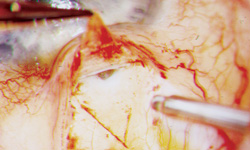
• Use of cautery. Underwater diathermy is a godsend for glaucoma surgeons. Bleeding is anathema, and underwater diathermy allows us to be very precise in eliminating it. Diathermy also avoids scarring and charring of the scleral tissue. Above all, we want to assiduously avoid having blood in the area of the filtration zone.
• Creating the sclerectomy. Location and size are important. The main thing is to position the sclerectomy anteriorly, so as to avoid iris incarceration or other obstructions postoperatively, and so that when you do your iridectomy, you avoid the peripheral iris vessels. Beyond that, you want to be uniform and consistent in the size of the sclerectomies you make. This will help you to know exactly what kind of IOP reduction will result.
When it comes to the scleral flap, the shape of the flap—triangle, rectangle or something else—probably doesn’t make too much difference. Most important are thickness, tightness of closure and posterior extent of the flap; these can affect scleral flap mobilization and outflow. For example, if the edge of your scleral flap is very close to the sclerectomy site, then when you do laser suture lysis or releasable sutures, the scleral flap will scroll up and there will be less tamponade. If the edge of the flap extends well past the sclerectomy, even if you do laser suture lysis there will be less release and elevation of the flap over the scleral flap.
• Consider using deep sclerectomy flap mobilization and unroofing Schlemm’s canal. When I do a routine trabeculectomy, I often create a sclerectomy flap underneath my traditional trabeculectomy flap, to unroof and expose Schlemm’s canal. Though not necessary in a standard trabeculectomy, I’m interested in the alternative Schlemm’s canal-based procedures such as canaloplasty and viscocanalostomy, and performing these steps allows me to improve my technique and become more comfortable with them. Doing this doesn’t compromise the trabeculectomy in any way; indeed, there are theoretical reasons it may enhance it by generating increased outflow through Schlemm’s canal.
• Use of antimetabolites. Advanced glaucomatous damage with significant visual field loss has been shown to require significant IOP lowering. To get a low pressure, we use broad application of an antimetabolite in a fixed dosage with a duration of up to three minutes or more. For other patients not requiring such a low pressure, the dosage or time of application might be reduced, or antimetabolites might not be used at all.
• Releasable sutures or laser suture lysis? Whether to use releasable sutures or use laser suture lysis is a matter of surgeon preference. I find them to be equally effective. I have a laser available to me in my clinic, so I favor laser suture lysis, but releasable sutures are also highly effective. They’re basically used in the same way as laser suture lysis.
The key thing is to use the tightness of closure to modulate flow. We titrate the flow on the table by deepening the anterior chamber with balanced salt solution and then assessing the flow underneath the flap. We’re striving for slow, steady egress of aqueous underneath the scleral flap while the anterior chamber is formed. We also may use digital massage and the Traverso maneuver, where we push on the side of the flap with a cotton-tipped applicator to test the flow.
• Positioning the sutures. The conjunctival sutures should be as far away from the action zone as possible. If I do a limbal-based flap, I want the incision to be as high up in the fornix as possible and as small as possible. If it’s a fornix-based flap, I like to use wing sutures of inert material that will not stimulate more scarring.
|
If possible, I favor scleral flap sutures parallel to the limbus as opposed to perpendicular to the limbus. Sutures that are perpendicular tend to create more astigmatism, which can affect visual recovery. This is particularly important for combined procedures.
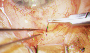
• Creating the peripheral iridectomy. I favor creating iridectomies in all phakic eyes. I like to have the peripheral iridectomy be broad and basal to avoid iris obstruction and incarceration of the sclerectomy postoperatively, and to minimize the chance of pupillary block. A PI may not be required in every combined glaucoma cataract operation, and we don’t do iridectomies when we use the ExPRESS shunt.
• Conjunctival closure. This is a complicated subject, in part because there are multiple closure techniques. Limbal-based flap closure can involve running and interrupted closure techniques that may or may not incorporate Tenon’s fascia. Fornix-based flaps closure techniques usually depend on whether you have retained a limbal remnant. If you do an anterior dissection of your fornix flap and do not leave a limbal remnant, most surgeons will use wing sutures; I often use horizontal mattress sutures in addition. If you’re leaving a limbal remnant, most surgeons will do a running closure of the conjunctival flap to the limbal remnant.
The take-home message here is that there are multiple ways to close conjunctiva. You need to develop a technique that works in your hands.
• Test for bleb leaks on the table. It’s very important to test for leaks intraoperatively and close them while you’re in the OR. Also, look for
leaks postoperatively using 2% fluorescein, and treat those appropriately.
At the Close of Surgery
For basic postoperative care, I routinely use topical cycloplegics to put the ciliary body at rest and to tighten the zonules to help maintain the anterior chamber depth.
We routinely have the patient use topical prednisolone acetate six times a day—more often in high-risk eyes. We also use a topical antibiotic for a couple of weeks.
|
In many cases, you’ll want to release the sutures to modulate the flow postoperatively. The big issue with either releasable sutures or laser suture lysis is the timing of release. If we’ve used intraoperative metabolites, we favor delaying the release of the sutures or performing laser suture lysis for at least a week postoperatively, to minimize the chance of converting a high pressure to a low pressure with a shallow chamber and choroidal attachment formation. If we didn’t use intraoperative metabolites, we prefer to do laser suture lysis or releasable sutures a little earlier; in that situation you can get scarring very quickly, and we want to enhance flow underneath the flap to elevate the bleb.
It’s a New Day
Having the ability to modify all of the factors described above allows us to choose and achieve a target pressure to a far greater extent than in the past. That’s very important, because glaucoma is not a single disease entity. Some patients truly need a pressure in the single digits, while others are fine with a pressure in the high teens. Depending on what the patient needs, we can now significantly modify our choice of operation and exactly how we execute it. REVIEW
Dr. Shingleton is an associate clinical professor of ophthalmology at Harvard Medical School and a clinical instructor at Tufts University School of Medicine. He has performed more than 50,000 cataract, glaucoma, surgical and laser operations.


