 |
| Some surgeons may have experienced instances in which the illumination gets dim during their surgeries and then brightens again. The idea behind using four LEDs to provide the illumination in the Proveo system is to avoid such fluctuations. |
Leica’s Proveo 8
Leica Microsystems’ newest microscope has two features designed to help the surgeon maintain good visualization during a procedure.
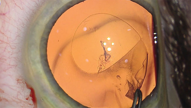
The first function is called CoAx 4, and is designed to use four LED light sources to keep the red reflex stable and optimize image contrast. Ike Ahmed, MD, assistant professor at the University of Toronto, has worked with the CoAx 4 system in the Proveo 8 both during its development and in its current form, and says it can be helpful in the OR. “The illumination and the stable red reflex are important features,” he says. “The red reflex is consistent throughout the procedure, during such maneuvers as the creation of the capsulorhexis; the red reflex isn’t necessarily better than in other microscopes, but it’s consistent. The microscope also maintains high-quality images throughout the case. For example, in the past, after hydrodissection, during phaco there’s a point at which we might lose the red reflex. With this system, however, we maintain good visualization throughout the steps of the procedure. I think this consistent visualization has to do with the way the coaxial light is delivered using the microscope’s four LEDs—there are no fiber optics involved in the system. By being a full-LED system, I think that leads to enhanced illumination when combined with the system’s optics. In the past, with some other scopes I might get great images at certain phases of the procedure but then lose them a bit, or I’d get unusual reflections that led to a loss of quality.” The system also allows the surgeon to adjust the diameter of the illumination from 4 to 23 mm, so he can illuminate just the iris or the entire eye. The idea is that a small diameter avoids scleral reflections and improves the contrast, while a larger diameter negates the negative effect of patient movement by keeping the diameter of light larger.
The other challenge faced by surgeons when using microscopes is the trade-off between depth of field and the amount of resolution of the image. To address this, the microscope uses a different aperture in each of the microscope’s ocular pathways. One aperture is larger and is designed for high-resolution imaging, while the other is smaller and geared toward providing a greater depth of field. The system then relies on the brain of the observer to fuse the disparate images into one that has both a high resolution and a good depth of field. “The enhanced depth of field, made possible by the asymmetrical apertures between the two oculars, is also really helpful,” says Dr. Ahmed. “This approach allows more things to be in focus at one time, which means less refocusing by the surgeon and better visualization across the various depths at which he’s working. During phaco, for instance, this comes into play when you’re bringing pieces of the nucleus up and down from the capsular bag to the iris plane.
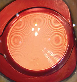 |
| The Proveo 8 system tries to provide a red reflex that remains consistent throughout the various phases of the procedure. |
| TrueVision Patents Its Tech |
| Recently, the U.S. Patent and Trademark Office awarded TrueVision 3D Surgical patents for digital surgical guidance as well as low-light imaging of surgical procedures. TrueVision’s surgical guidance and visualization patent describes a visualization platform that uses computer-guidance for different steps of ocular surgery, including aiding with the creation of a capsulorhexis and the centering of a corneal inlay for presbyopia treatment. The surgeon can make real-time adjustments to his inputs, such as changing the selected lens, and the system will change its guidance to allow him to reach the postop target refraction. The low-light imaging patent describes a system that would enable surgeons to visualize target tissues and structures accurately with the use of very little illumination. The company says surgeons may be able to dramatically reduce the use of the microscope light during procedures, which can be especially useful in vitreoretinal work. |
For information, call 1 (800) 248-0123, or visit leica-microsystems.com.
Topcon HD Video System
Topcon has released a modular high-definition video system to let surgeons capture procedures in higher detail.
“Recording in higher resolution lets the surgeon capture very small details,” says Topcon’s Ricardo Almiron. “The details are important when you’re dealing with sutures, IOL haptics or when a surgeon is performing retinal procedures such as peeling a subretinal membrane. Having these details is helpful when teaching or projecting an image for the surgeon’s assistants or out to an operating room theater.”
The base HD video system, which is a Panasonic product, has four components: a camera; camera control unit; cable; and a power supply. The camera attaches to the surgical microscope’s beam splitter. The system is described as fitting Topcon microscopes; however Mr. Almiron says it can fit most microscopes. Though the camera is Panasonic, Topcon provides the video adaptor that connects it to the microscope, and optimizes the color balance for ocular surgery.
Since various facilities may already have complementary components such as HD recorders and monitors, those are sold separately. “Facilities such as hospitals already have video monitors attached to booms affixed to the ceiling or the wall,” Mr. Almiron explains. “But if a facility needs it, we also sell a 26-inch HD monitor. We also make available a recorder that records to an SD card. It comes with 32 GB of memory, but that can be expanded to 64 GB, and it will allow up to 12 hours of recording depending on the resolution the surgeon chooses.”
The price for the HD video system is $7,675, though a discount is available for Veterans Administration facilities. The optional HD monitor is $7,995 and the recorder sells for $5,970. For information, call 1 (800) 223-1130, or visit topconmedical.com. REVIEW
Dr. Ahmed is a consultant for Leica.