Alcon’s Applied Integration
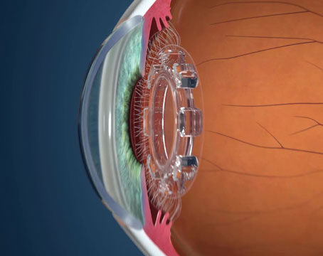
Alcon’s Cataract Refractive Suite connects an imaging/measurement device called the Verion with the Centurion phaco machine, the LenSx femtosecond cataract laser and the Luxor operating microscope.
“The goal is to have all of the surgeon’s in-office data acquisition easily transferable to the OR suite,’” says Richard J. Mackool Jr., MD, of Astoria, N.Y., who uses the system. “In the past, we would do the testing, obtaining the IOL power and astigmatism results on paper, and then make decisions about our lens choice in the OR based on those measurements. With the Verion, the data is stored digitally on a USB memory stick that is taken to the OR and inserted into the system. The microscope retrieves the data, giving you a digital overlay that’s visible in the microscope oculars and indicates the correct axis at which to implant the toric IOL. Before this, we’d mark the eye with a pen with the patient sitting up to indicate the principal meridians, then use a toric axis marker to locate the desired meridian. With the Verion, these steps are eliminated. Instead, you have a digital, real-time overlay as a direct indicator of the preop data.”
|
For lens selection, the Verion has such formulas as the Holladay, Holladay II, SRK/T and the Hoffer Q. “More important, after you’ve done a number of cases and entered the results into the system, the Verion will optimize your case results in the future by retrospectively analyzing the data so you can tailor the program you choose to the cases you perform. For instance, everyone has a different surgical technique: Some utilize a larger capsulorhexis than others, and the effective lens position of the IOL may be different in their patients than it would be for surgeons who make a smaller capsulorhexis.”
If a surgeon uses a LenSx femtosecond laser, Dr. Mackool says the Verion data is transferrable to it as well. “The same information provided to the OR for toric lenses can be imported into the LenSx for creating arcuate incisions,” he says. “Some surgeons prefer arcuate incisions, or combine them with a toric lens. Either way, the axis goes directly to the LenSx.”
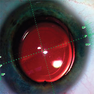
The other part of the equation, the Luxor operating microscope, also has features to aid the surgery. “Regardless of whether the patient is looking at the microscope or looking away, the red reflex is extremely well-maintained,” says Richard Mackool Sr., MD, who also uses the system. “As such, areas of the red reflex never become dark, even in pretty extreme directions of gaze on the patient’s part. This comes in handy if you want to rotate the globe a bit in a certain direction to gain access to one area or another, which happens all the time during surgery. In fact, the reflex is so good that we rarely need to use more than 50 to 60 percent of maximum illumination.”
For information on the Verion and the integrated system, visit https://www.myalcon.com/products/surgical/verion-guided-system/.
The Zeiss Cataract Suite
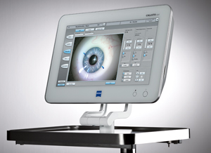
The brains of the integrated cataract suite from Carl Zeiss Meditec is the new Callisto computer-assisted surgery system, which acts as a hub for the company’s IOLMaster device and the OPMI Lumera microscope.
Richard Davidson, MD, associate professor of ophthalmology and medical director for the faculty practice at the University of Colorado Hospital Eye Center, used the Callisto system in its prototype stage for capsulotomy creation and the placement of toric lenses, and says he sees the potential of such integrated systems. He has also worked with the Alcon system. “This is the direction cataract surgery is heading—the integrated system,” he says. “The main benefits to the surgeon are convenience and patient safety. If you take the measurements for the IOL in your office and then transfer them to the machine, either the femtosecond laser or straight to the operating room microscope, you don’t have to worry about paper charts getting lost, choosing the wrong lens, or the lens not matching up correctly with the patient.”
|
If the surgeon upgrades to Callisto Eye Assistance, the Callisto display will show several forms of digital overlays to guide the physician during different phases of the procedure. If the Integrated Data Injection System is also installed, these overlays will appear right in the microscope. There are currently guides for corneal incisions, including limbal relaxing incisions; capsulorhexis creation; Z Align for help in aligning toric IOLs; and K Track, which helps visualize corneal curvature in combination with a keratoscope. “For now, you have to mark the eye preoperatively and the system tracks your marks,” says Dr. Davidson. “But the ultimate goal is to track based on a preop eye image. When the guides are on, the limbus is marked so you can always see it, and you also have marks for the main axis and the axis 90 degrees away from it. You get a pretty precise idea of where the lens needs to go.”
The other eventual goal of the system is to be able to transfer the information wirelessly between devices, or at least over a network. “When I used the system, the data was transferred with a flash drive,” says Dr. Davidson. “Our hospital system here is complex, so getting access to the network can be difficult, so we just used a flash drive. Eventually it will be networked and, hopefully, wireless as well. The plan is, if you have a surgery center in your office, to be able to do your IOLMaster and then walk into the OR and turn on the Callisto machine and see all of your data right in front of you.”
For information on Zeiss’ integrated cataract system, visit http://meditec.zeiss.com/meditec/en_us/products.html. REVIEW