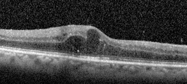
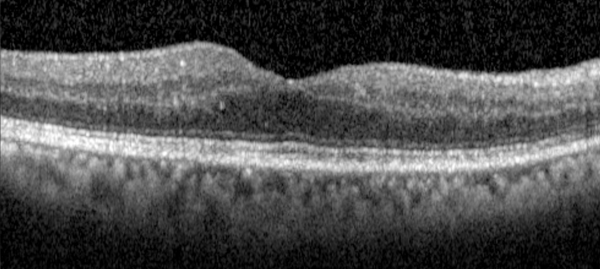
Clinically significant diabetic macular edema has long been recognized as a major cause of loss of vision in patients with diabetic retinopathy.1,2 As the prevalence of diabetes grows worldwide, the potential loss of vision from DME poses a significant concern with regard to quality of life and socioeconomic considerations.3,4
The treatment for DME with focal/grid laser to microaneurysms or areas of diabetic macular edema has been established since the Early Treatment Diabetic Retinopathy Study.5 However, when the Diabetes Retinopathy Clinical Research Network (DRCRnet) published a prospective trial comparing standard laser therapy to combination therapy with ranibizumab or steroid, the standard of care became challenged, as the combination therapy group (laser and ranibizumab) demonstrated superior visual outcomes in the treatment of DME when compared to laser monotherapy.6
Following this promising result, the RESTORE trial was the first large randomized clinical trial to evaluate if ranibizumab alone or in combination with laser was more efficacious than macular laser alone.9 The trial demonstrated that ranibizumab as monotherapy or in combination with focal/grid macular laser provided superior visual acuity outcomes over focal/grid macular laser alone in patients with DME. At one year, no differences were detected between the ranibizumab and ranibizumab/laser arms, and approximately seven injections were necessary in the ranibizumab arms. The two-year safety and efficacy data were presented for the RESTORE Extension Study (Mitchell P. IOVS 2012;53:ARVO E-Abstract 4667). Patients were treated with ranibizumab on an “as needed” basis and/or with laser. Retreatment occurred if there was a decrease in BCVA due to DME progression, confirmed by clinical evaluation and/or OCT or in the opinion of the investigator.9 The gains in BCVA that were observed in the first 12 months were maintained at month 24. There were no safety signals noted in either arm. During year two, approximately 3.9 injections were necessary in the ranibizumab monotherapy arm versus 3.5 in the laser/ranibizumab arm. The addition of laser did not significantly lessen the burden of ranibizumab injection therapy.
The REVEAL trial had a design similar to that of the RESTORE trial, except that it followed an Asian cohort with DME (Ohji M, et al. IOVS 2012;53:ARVO E-Abstract 4664). At one year, the REVEAL trial also demonstrated the superior outcomes of ranibizumab and ranibizumab with laser (+5.9/+5.7 ETDRS letters gained) versus laser monotherapy (+1.4 ETDRS letter gained). The mean number of injection treatments was 7.8 in the ranibizumab monotherapy vs. 7.4 in the ranibizumab/laser combo therapy. There were no significant safety signals (ocular or systemic) noted in this trial.
Ranibizumab Monotherapy & DME
For many years it has been established that vascular endothelial growth factor plays a role in the creation of retinal ischemia and increased vascular permeability that gives rise to macular edema.7,8 The superiority of ranibizumab monotherapy over laser alone has been noted.9 The RISE and RIDE trials were identical, double-masked, sham-controlled, multicenter Phase III trials evaluating the impact of monthly ranibizumab injections on DME (Ip M, et al. IOVS 2012;53:ARVO E-Abstract 1336).12 Both trials evaluated sham vs. 0.3-mg vs. 0.5-mg ranibizumab monthly monotherapy in the treatment of DME over a 24-month time frame, with additional treatment and follow-up out through 36 months. After three months of injection therapy in the trial, rescue macular laser could be applied if it were found that central foveal thickness was >250 µm or if there was a 50-µm worsening from the prior month.
For the RISE trial, 377 patients were randomized (127 to sham, 125 to 0.3 mg, 125 to 0.5 mg) with the characteristics similar across the three arms. At 24 months for >15 letter visions gains, 18.1 percent of sham patients versus 44.8 percent of 0.3-mg and 39.2 percent of 0.5-mg ranibizumab patients were noted. In theRIDE study, 382 patients were randomized (130 to sham, 125 to 0.3 mg, 127 to 0.5 mg) with similar baseline characteristics. For the proportion of patients experiencing >15 letter vision gains, 12.3 percent of sham patients versus 33.6 percent of 0.3-mg versus 45.7 percent of 0.5-mg ranibizumab patients were noted. Pooling the efficacy data, the visual results, both the proportion of eyes gaining three or more lines and mean BCVA, were identical for the two doses. Significant improvements in macular edema were noted on OCT in both ranibizumab arms in both trials; retinopathy was less likely to worsen in ranibizumab-treated patients.
|
Both RISE and RIDE established ranibizumab monthly monotherapy as an efficacious and sustainable treatment for DME, with low rates of ocular and systemic complications for up to 36 months. In August 2012, ranibizumab at the 0.3-mg dose gained Food and Drug Administration approval for the treatment of DME in the United States.
Alternative Treatment Prospects
Besides targeting VEGF with ranibizumab, there are other promising treatment modalities that may offer additional help for the treatment of DME.
Bevacizumab has an established treatment history for macular degeneration and DME. Its use in ophthalmology remains off-label; however, in ophthalmology it is a widely accepted treatment for exudative age-related macular degeneration.13 Its application to treat DME or proliferative diabetic retinopathy has an extensive clinical history, although long-term, prospective, comparative clinical trial data is limited.
In DME, bevacizumab has been evaluated in a prospective study, the BOLT trial.14 This study consisted of 80 patients that were randomized to 1.25-mg bevacizumab versus standard macular laser for non-ischemic, center-involving, clinically significant macular edema. At 12 months, bevacizumab led to significant gains in ETDRS letters versus laser monotherapy (median gain of eight ETDRS letters vs. median loss of 0.5 ETDRS letters in the laser group).14 The application of bevacizumab is a reasonable alternative treatment for DME at the current time.
Similar to bevacizumab, intravitreal triamcinolone for DME also remains an off-label application. In the DRCRnet trial, the triamcinolone with laser therapy arm was found to have visual gains when evaluated in pseudophakic patients.6 However, the possibility of provoking cataract formation or the potential for steroid-induced ocular hypertension or the possible exacerbation of glaucoma have placed its use secondary to anti-VEGF-based strategies.
Future Therapeutic Prospects
Ozurdex is a sustained-release dexamethasone intravitreal office-based injectable implant that has FDA approval for the treatment of branch and central retinal vein occlusion-associated macular edema and for the treatment of posterior non-infectious uveitis. Its use for DME remains off-label, but promising.
One such study evaluated persistent DME >90 days to one of two intravitreal dexamethasone implant doses (350 micrograms or 700 µg) versus observation.15 The study evaluated 171 eyes; at day 180 best-corrected visual acuity improvement of 10 letters or more was seen in 30 percent of eyes in the 700-µg group, 19 percent in the 350-µg group, and 23 percent in the observation group (p≥ 0.4 for treated vs. observed eyes). There were also significantly greater improvements in central retinal thickness and fluorescein leakage. Another study evaluated the efficacy of Ozurdex in refractory DME in post-vitrectomy eyes and found improved vision and OCT-determined central thickness with the 700-µg implant.16
Iluvien is another promising sustained-release steroid, intravitreal, office-based implant that utilizes fluocinolone as opposed to dexamethasone. The advantages of this particular platform include a smaller size (25 ga. as opposed to 22 ga. with Ozurdex) and a longer duration of efficacy (2.5 to three years).
FAME, a prospective, randomized trial, just published its three-year data.17 The trial evaluated two different doses of steroid implant (0.2 µg /day versus 0.5 µg /day) versus sham control. At three years, the percentage of >15 letters of vision gained was 28.7 percent (0.2 µg /day) and 27.8 percent (0.5 µg /day) in the implant groups compared with 18.9 percent (p=0.018) in the sham group. Virtually all phakic patients developed cataracts, but their visual benefit after cataract removal was similar to that of patients who were pseudophakic at baseline. The incidence of incisional glaucoma surgery was found to be 4.8 percent in the low-dose group and 8.1 percent in the high-dose insert group. Iluvien is approved for treating DME in Europe but not in the United States.
Aflibercept (Eyelea) is a commercially available drug that is FDA-approved for the treatment of exudative AMD. Its role in the treatment of DME is promising and currently undergoing Phase III testing for this indication. Aflibercept is a recombinant fusion protein comprising the key VEGF-binding domains of human VEGF receptors 1 and 2 with a higher binding affinity versus ranibizumab and bevacizumab, along with binding capacity for placental growth factor, which has been shown to contribute to excessive vascular permeability and retinal neovascularization.18,19 The Phase II experience of aflibercept for treating DME (DA VINCI trial) was recently published.20 Two hundred twenty-one patients with center-involving DME were randomized to one of five treatment regimens: aflibercept 0.5 mg every four weeks; 2 mg every four weeks; 2 mg every eight weeks after three initial monthly doses; 2 mg dosing as needed after three initial monthly doses; or macular laser photocoagulation. The primary outcomes were BCVA at 24 weeks and at 52 weeks, proportion of eyes that gained 15 or more letters in ETDRS BCVA, and the mean changes in central foveal thickness as assessed by OCT. At 52 weeks, the mean improvements in BCVA in the respective aflibercept groups were 11, 13.1, 9.7 and 12 letters versus, 1.3 letters for the laser group. The proportions of eyes obtaining >15 ETDRS letters were 40.9 percent, 45.5 percent, 23.8 percent and 42.2 percent versus 11.4 percent for laser. The mean reduction in central foveal thickness by OCT for the aflibercept groups were 165.4 µm, 227.4 µm, 187.8 µm and 180.3 µm versus 58.4 µm for laser. There were no significant ocular or systemic safety signals identified in the trial.
The Evolving Landscape
It is striking to think that, with more than 20 years of treatment for DME, macular focal/grid laser monotherapy is now called into question as the best clinical practice. There is now enough separate and repeated level-one evidence available to change the standard of care for treating center-involving DME to anti-VEGF therapy, particularly ranibizumab with or without laser treatment. Although we know that adding laser to anti-VEGF therapy doesn’t provide better visual outcomes, it may decrease total injections in some patients over the long run. The exact best injection treatment protocol has yet to be established (i.e., monthly injection versus as-needed versus “treat and extend”). An interesting result from the RISE and RIDE studies is the suggestion that ranibizumab monotherapy had a lower progression to proliferative retinopathy than the sham arm. Bevacizumab and triamcinolone remain readily available alternatives to ranibizumab.
For initial DME with decreased baseline visual acuity, the existing published data currently favors an anti-VEGF strategy over a steroid-based therapy as initial therapy, especially for phakic eyes. The role of aflibercept has yet to be established but its Phase II data demonstrates promising efficacy and safety. The extended-release steroid devices are intriguing for post-vitrectomized eyes or potentially for resistant cases of DME, and future studies and data will be welcome. Analogous to the shift in therapy for AMD with the ANCHOR and MARINA trials in 2006,12,13 we are now seeing the landscape change with our approach to DME. REVIEW
Dr. Shah is a fellow in the Retina Service at Wills Eye Institute. Dr. Regillo is the director of the Retina Service at Wills, a professor of ophthalmology at Thomas Jefferson University, and a partner at Mid Atlantic Retina.
1. Klein R, Klein BE, Moss SE, et al. The Wisconsin Epidemiologic Study of Diabetic Retinopathy. IV: Diabetic macular edema. Ophthalmology 1984;91:1464-74.
2. MossSE, KleinR, KleinBE. The 14-year incidence of visual loss in a diabetic population. Ophthalmology 1998;105:998-1003.
3. International Diabetes Federation. IDF Diabetes Atlas, 4th ed. Brussels, Belgium: IDF Executive Office; 2009. Available at: http://www.diabetesatlas.org/. Accessed April 20, 2011.
4. Javitt JC, Aiello LP, Chiang Y, et al. Preventive eye care in people with diabetes is cost-saving to the federal government: implications for health-care reform. Diabetes Care 1994;17:909-17.
5. Early Treatment Diabetic Retinopathy Study Research Group. Photocoagulation for diabetic macular edema: Early Treatment Diabetic Retinopathy Study report number 1. Arch Ophthalmol 1985;103:1796-806.
6. Diabetic Retinopathy Clinical Research Network; Elman MJ, Aiello LP, Beck RW, et al. Randomized trial evaluating Ranibizumab plus prompt or deferred laser or triamcinolone plus prompt laser for diabetic macular edema. Ophthalmology 2010;117:1064-77.
7. Cunha-Vaz J, Faria de Abreu JR, Campos AJ. Early break- down of the blood-retinal barrier in diabetes. Br J Ophthalmol 1975;59:649-56.
8. Qaum T, Xu Q, Joussen AM, et al. VEGF-initiated blood-retinal barrier breakdown in early diabetes. Invest Ophthalmol Vis Sci 2001;42:2408-13.
9. Mitchell P, Bandello F, Schmidt-Erfurth U, et al, RESTORE Study Group. The RESTORE Study: Ranibizumab monotherapy or combined with laser versus laser mono-therapy for diabetic macular edema. Ophthalmology 2011;118:615-25.
10. Brown DM, Kaiser PK, Michels M, et al. Ranibizumab versus verteporfin for neovascular age-related macular degeneration. N Engl J Med 2006;355:1432-1444.
11. Brown DM, Michels M, Kaiser PK, et al. Ranibizumab versus verteporfin photodynamic therapy for neovascular age-related macular degeneration: Two-year results of the ANCHOR study. ANCHOR Study Group. Ophthalmology 2009;116:57-65.
12. Nguyen Q, Brown D, Marcus D. Ranibizumab for Diabetic Macular Edema Results from 2 Phase III Randomized Trials: RISE and RIDE. Ophthalmology 2012;119:789-801
13. Martin DF, Maguire MG, Ying GS, et al. Ranibizumab and bevacizumab for neovascular age-related macular degeneration. N Engl J Med 2011;364(20):1897-1908.
14. Michaelides M, Kaines A, Hamilton R, et al. A Prospective Randomized Trial of Intravitreal Bevacizumab or Laser Therapy in the Management of Diabetic Macular Edema (BOLT Study) Ophthalmology 2010;117:1078-1086.
15. Haller J, Kuppermann B, Blumenkranz M. Randomized Controlled Trial of an Intravitreous Dexamethasone Drug Delivery System in Patients With Diabetic Macular Edema. Arch Ophthalmol 2010;128:289-296.
16. Boyer D, Faber D, Gupta S, et al. Dexamethasone Intravitreal Implant for Treatment of Diabetic Macular Edema in Vitrectomized Patients. Retina 2011;31:915-923.
17. Campochiaro P, Brown D, Pearson A, et al. Sustained Delivery Fluocinolone Acetonide Vitreous Inserts Provide Benefit for at Least 3 Years in Patients with Diabetic Macular Edema. Ophthalmology 2012 Article in Press.
18. Holash J, Davis S, and Papadopoulos N, et al. VEGF-Trap: A VEGF blocker with potent antitumor effects. Proc Natl Acad Sci USA 2002;99:11393-8.
19. Rakic JM, Lambert V, Devy L, et al. Placental growth factor, a member of the VEGF family, contributes to the development of choroidal neovascularization. Invest Ophthalmol Vis Sci 2003;44:3186-93.
20. Do D, Nguyen Q, Boyer D, et al. One-Year Outcomes of the DA VINCI Study of VEGF Trap-Eye in Eyes with Diabetic Macular Edema. Ophthalmology 2012 online Article in Press.


