Presentation
A 74-year-old Caucasian man presented for a second opinion regarding a recurrent ocular surface lesion of the left eye. Four years prior to presentation he was treated for a “squamous papilloma” of the conjunctiva with excision and subconjunctival interferon (IFN). The lesion subsequently recurred three times. The patient underwent re-excision with postoperative topical mitomycin C after the first recurrence, then a second re-excision and treatment with topical 5-fluorouracil. When he developed a third recurrence, proton beam radiation was recommended at which time he sought a second opinion at the Wills Eye Hospital Oncology Service.
Medical History
Past ocular history included cataract surgery in both eyes, ocular hypertension and dry age-related macular degeneration. Past medical history included hyperlipidemia. Family history disclosed a sister with primary open-angle glaucoma. Social history was non-contributory. Current medications were travoprost, AREDS2 vitamins and simvastatin.
Examination
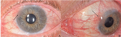 |
| Figure 1. External photographs of the left eye in primary gaze (A) and abduction (B) upon presentation to Wills Eye Hospital. A 4 x 4 mm fibrovascular frond extends from the conjunctiva onto the corneal surface at the superonasal limbus (arrows). |
On examination, visual acuity with correction was 20/30 OD and 20/60 (pinhole 20/50) OS. Pupils were normal, and intraocular pressures were 17 mmHg OD and 18 mmHg OS. Extraocular movements and confrontation visual fields were full in both eyes.
Anterior segment examination of the left eye revealed a 4 mm x 4 mm fibrovascular frond at the superonasal limbus extending onto the cornea (Figure 1). Additionally, there was a posterior chamber intraocular lens in both eyes. Dilated fundus examination demonstrated a few macular drusen but was otherwise unremarkable.
What is your diagnosis? What further workup would you pursue? Click here for the diagnosis.


