A 25-year-old man who
 |
Medical History
The patient’s ocular history was notable only for trauma to the right eye from a piece of plastic which had left a partial thickness corneal laceration inferotemporally several years prior. He had not had any previous eye surgeries. He denied any past medical history or current medication use. He had no allergies to any medications and his family and social histories were noncontributory.
Examination
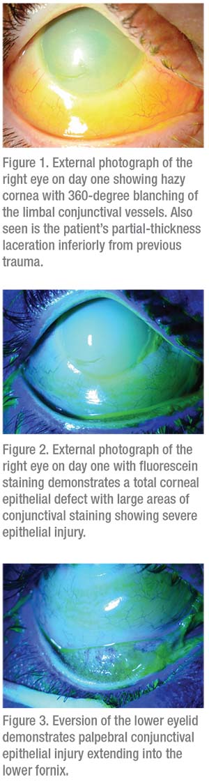
Ophthalmic examination revealed uncorrected Snellen visual acuity of 20/200 in the right eye with no improvement with pinhole, and 20/25 in the left eye. His right pupil was difficult to examine but was seen to be somewhat reactive in the slit lamp. The left pupil was briskly round and reactive. No afferent pupillary defect was present. Extraocular movement and confrontational visual fields were full in both eyes. Intraocular pressures were measured by applanation at 16 mmHg in the right eye and 18 mmHg in the left eye. External and anterior segment examination revealed ptosis of the right eye with moderate upper and lower eyelid edema and erythema. Examination of the conjunctiva revealed chemosis inferiorly with 360-degree blanching of the limbal conjunctival vessels and an almost 100-percent conjunctival epithelial defect (Figures 1-3). The cornea was diffusely whitened with a total epithelial defect and a partial-thickness laceration inferotemporally from the previous trauma. There was a poor view to the iris through the hazy cornea but it was seen to be flat and the pupil round, and the anterior chamber was deep. There was a hazy view to the lens but no gross abnormality was seen. The anterior examination of the left eye was normal. B-scan of the right eye demonstrated a formed anterior chamber with the lens in place, clear vitreous and a flat retina.
Click here for workup, diagnosis and discussion.


