Ocular surgery, whether cataract, cornea, glaucoma or retina has evolved to become highly efficient and predictable. However, complications can still occur, including corneal edema and decompensation, lens subluxation, vitreous loss, macular edema and retinal detachment. Eyelid malposition can occur after any ocular surgery including ectropion,1 entropion2 and ptosis. The incidence of ptosis after cataract surgery has been reported to be as high as 13 percent3; the rate of entropion and ectropion after cataract surgery is not known.
The cause of postoperative ptosis is manifold including postoperative edema, anesthesia and surgical technique. This article will discuss the evaluation, etiology, treatment and prevention of ptosis after ocular surgery.
Classification of Ptosis
Ptosis can be divided into its etiologies, including myogenic, aponeurotic, neurogenic, mechanical or traumatic.4
Myogenic ptosis is typically caused by congenital dysgenesis of the levator muscle or an acquired muscular dystrophy. In post-surgical myogenic ptosis, direct damage to the levator muscle may be due to the process of injecting anesthetic into the muscle or myotoxic effects of the anesthesia.
Aponeurotic ptosis is a disinsertion or dehiscence of the levator aponeurosis from its normal position on the anterior surface of tarsus. The use of a bridle suture or rigid lid speculum has been implicated in cataract surgery as a cause of aponeurotic damage.
Neurogenic ptosis is a disruption of the innervation of the muscle, and in the case of post-surgical ptosis, the prolonged effects of anesthetic on the neuromuscular junction cause this transient phenomenon. This may also be caused by susceptibility of anteriorly located terminal twigs of the oculomotor nerve to local anesthesia infiltrated in the eyelid in a Van Lint block.5
Mechanical ptosis occurs when a mass causes downward pull on the upper eyelid. In postoperative ptosis this may be due to edema or hematoma formation in the eyelid. Finally, traumatic ptosis is due to blunt or sharp trauma to the levator aponeurosis. All of the causes discussed in the categories above can be included under the heading of trauma.
Post-surgical ptosis is an acquired ptosis in which duration of the ptosis can help determine the etiology. Ptosis that resolves after surgery is considered transient or acute ptosis; ptosis that persists after surgery is categorized as chronic or persistent.
Transient Ptosis
Transient ptosis, or ptosis that improves over the postoperative period, can be caused by eyelid edema, hematoma formation (both intraorbital and eyelid), foreign body reaction, ocular inflammation and anesthesia effects.
 |
Hematoma formation, usually secondary to local injection of the eyelids during a Van Lint block, is a form of mechanical ptosis. However, even intraorbital hematoma secondary to peribulbar or retrobulbar anesthesia can cause limited function of the levator muscle and secondary ptosis. Ocular massage or compression after injection can limit hematoma formation.10 Resorption of a hematoma can cause fibrosis and adhesions between the orbital septum and levator aponeurosis and create persistent ptosis.
Foreign-body reaction is overlooked as a cause of ptosis. In extracapsular cataract surgery, nylon sutures can erode through overlying conjunctiva and cause inflammation and edema in the eyelid.11 Ocular inflammation and surface irritation after surgery also create a transient, reactive ptosis. With correction of the inflammation, the edema and ptosis resolve.
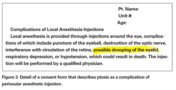
Short, transient ptosis can occur as a direct response to the neuromuscular blockade of the levator muscle. This can be secondary to a retrobulbar or peribulbar block with anesthetic bathing the levator complex. Furthermore, lid infiltration can affect distal fibers of the oculomotor nerve. Choice of anesthetic can determine the duration of ptosis, lidocaine having a shorter duration than bupivicaine.
The use of hyaluronidase can also alter the effects of local anesthetic; it acts to increase the spread of anesthetic between tissue planes by action of hyaluronic acid.12 No study has directly evaluated the effects of hyaluronidase on postoperative ptosis, but increased availability of anesthetic may increase the incidence of ptosis. Adjusting pH of the anesthetic can also increase its availability; buffering an anesthetic with bicarbonate can increase the pH of local anesthetic. Compared to hyaluronidase, anesthetic buffered with bicarbonate had a higher rate of ptosis in one study. This may be due to increased availability of anesthetic with buffering or increased toxicity due to the different pH.
Finally, ocular massage or compression can increase availability of anesthetic. Although it has been demonstrated to decrease lid edema and hematoma formation, increased availability of anesthesia in the peribulbar or retrobulbar space can increase the rate of ptosis.
If ptosis persists past the neuromuscular effects of the anesthetic, one must suspect the myotoxic effects of anesthesia. One study demonstrated that lidocaine causes degeneration of human muscle 18 hours after injection.13 Epinephrine heightens the myotoxic effects of local anesthetic.14 Recovery of muscle function and resolution of ptosis occur both due to regeneration of damaged muscle fibers, and hypertrophy of remaining muscle fibers15; this process should take between eight and 12 weeks.16
Persistent Ptosis
Causes of persistent or chronic ptosis usually involve damage to the levator aponeurosis or scarring to the levator complex. Direct damage to the muscle via toxic effects of anesthesia or trauma from direct injection of the muscle usually resolves with regeneration of the muscle. Dehiscence or detachment of the levator aponeurosis from the tarsal plate does not remedy itself; therefore, this form of ptosis does not improve over the postoperative period.
 |
The use of a bridle suture during cataract surgery has also been implicated in post-surgical ptosis. Ptosis occurs both by grasping the superior rectus during passage of the bridle suture and traction of the superior rectus by the bridle suture. Michael Loeffler, MD, and colleagues demonstrated that open visualization of the superior rectus, versus blind passage of the bridle suture by grasping the superior rectus with forceps, decreases the amount of postoperative ptosis.17 The effect of the bridle suture is enhanced with the use of a lid speculum. Traction on the superior rectus/levator complex while the upper lid is rigidly fixated with a speculum can cause marked dehiscence of the aponeurosis.3 Jignesh Patel and colleagues, however, found no significant difference of ptosis development in patients who had a bridle suture placed versus those who did not.18
Finally, the lid speculum has been indicated as a cause of persistent ptosis. The effects of the lid speculum are not dependent on the use of a bridle suture. John Linberg, MD, and associates demonstrated ptosis after refractive surgery where a bridle suture is not used.19 The authors speculate that use of a rigid speculum versus a flexible wire speculum may cause dehiscence of the aponeurosis when the lid is forcefully squeezed or blinked; a wire speculum will yield to such forces.
Intervention
After a thorough examination in which the etiology is determined, one must decide whether to intervene. In most cases, post-surgical ptosis resolves with time, and therefore observation is the most prudent form of intervention. As in other forms of traumatic ptosis, this form of ptosis typically improves within six months. Fereydoun Parsa, MD, and associates reported a case of spontaneous resolution of ptosis after 11 months and suggest observing post-surgical ptosis for one year.20
Ptosis that does not resolve is typically secondary to aponeurotic dehiscence; this is readily repaired surgically. However, prior to considering surgical intervention, one must determine whether the patient is affected by the ptosis. In one study, only 18 percent of patients with postoperative ptosis noticed a change in their eyelid postion.8 Finally, plans for contralateral ocular surgery have to be determined. If a patient is planning ocular surgery in the other eye, surgical intervention for ptosis should be delayed, since the second eyelid is at similar risk for postoperative ptosis.
Ptosis after ocular surgery is amenable to repair via an external approach through the lid crease and repair of the aponeurotic dehiscence or a transconjunctival approach with Müellerectomy for minimal ptosis. The former approach is more logical since it directly addresses the dehiscence of the levator aponeurosis. Furthermore, patients with a dehisced levator will have an increased lid crease height and a superior sulcus defect secondary to retraction of the preaponeurotic fat pad. External levator resection through the anatomic lid crease addresses all these problems; an internal approach does not. However, for minimal ptosis of 1 to 2 mm, a Müellerectomy may be appropriate.
Prevention
Prevention of post-surgical ptosis is an essential part of modern ocular surgery. Even if a patient obtains excellent visual acuity from cataract surgery, a patient's function may be limited by post-surgical ptosis. Therefore, a surgeon should take an active role in preventing this problem.
 |
Surgical technique can also determine exposure to risk factors for post-operative ptosis. An efficient surgeon can limit surgical time and thus eyelid complications secondary to ocular inflammation or compressive effects of prolonged use of a lid speculum. Also, disuse of bridle sutures or a rigid speculum will limit these factors in ptosis. If a bridal suture is necessary for fixation of the globe, direct visualization of the superior rectus with dissection through conjunctiva and Tenon's capsule during placement of the bridle suture will limit damage to the levator complex. An episcleral or corneal traction suture may also be considered. Use of a flexible wire speculum and limiting the spreading tension on the eyelids may also limit postoperative lid malposition. If a rigid speculum is required, lid akinesia with anesthesia might limit aponeurotic damage.
Surgical technique may also decrease the incidence of ptosis; one study found a higher incidence of ptosis in extracapsular cataract extraction versus phacoemulsification at eight weeks.6 However, there was no difference at six months after surgery. This difference may be due to a larger conjunctival flap and scleral incision, as well as more sutures needed in wound closure, increasing the likelihood of postop inflammation. With the advent of temporal, clear corneal surgery, these factors are reduced. One can also speculate that the superior approach to surgery may have a greater risk of ptosis compared to a temporal approach, since vertical traction of the globe during a superior approach may cause dehiscence of the aponeurosis, whereas a temporal approach has no vertical vectors applied to the globe.
Lid malposition after ocular surgery should be recognized as a preventable complication. Understanding the causes of eyelid malposition after surgery can help a surgeon prevent its occurrence with careful surgical planning. When present after surgery, a thorough examination can help determine when, and if, surgical intervention is indicated. Finally, patients should be educated about its possibility as a complication in routine ocular surgery.
Drs. Crum and Bernardino are at the Yale School of Medicine, Department of Ophthalmology, Section of Ophthalmic Plastics and Orbital Surgery. Contact Dr. Bernardino at Yale Eye Center, 40 Temple Street, 3D, New Haven, Conn. 06510. Phone: (203) 785-2020; fax: (203) 785-5909; e-mail:
crbernardino@mac.com.
1. Hosal BM, Gürsel TE. Eyelid malpositions after cataract surgery. Euro J Ophthalmol 1998;8:12-15.
2. Hurwitz JJ, Smith D, Corin SM. Association of entropion with cataract surgery. Ophth Plast Reconstr Surg 1990;6:25-27.
3. Alpar JJ. Acquired ptosis following cataract and glaucoma surgery. Glaucoma 1982;466-68.
4. Beard C. Ptosis, 3rd ed. St. Louis: Mosby, 1981.
5. Hwang K, Lee DK, Chung IH, Lee SI. Patterns of oculomotor nerve distribution to the levator palpebrae superioris muscle, and correlation to temporary ptosis after blepharoplasty. Ann Plast Surg 2001;47:381-384.
6. Kaplan LJ, Jaffe NS, Clayman HM. Ptosis and cataract surgery. A multivariant computer analysis of a prospective study. Ophthalmol 1985;92:237-242.
7. Wolfort FG, Poblete JVP. Ptosis after blepharoplasty. Ann Plast Surg 1995;34:264-267.
8. Feibel RM, Custer PL, Gordon MO. Postcataract ptosis. A randomized, double-masked comparison of peribulbar and retrobulbar anesthesia. Ophthalmol 1993;100:660-665.
9. Paris GL, Quickert MH. Disinsertion of the aponeurosis of the levator palpebrae superioris muscle after cataract extraction. Am J Ophthalmol 1976;81:337-340.
10. Ropo A, Ruusuvaara R, Paloheimo M, Maunuksela EL, Mikki P. Periocular anaesthesia: Technique, effectiveness and complications with special reference to postoperative ptosis. Acta Ophthalmologica 1990;68:728-732.
11. Shahinian L, Brown SI. Postoperative complications with protruding monofilament nylon sutures. Am J Ophthalmol 1977;83:546-548.
12. Mather C, Smith JH, Bloom PA. The efficacy of 0.75% bupivicaine with ph adjustment and hyaluronidase for peribulbar blockade: the incidence of prolonged ptosis. Euro J Ophthalmol 1994;4:13-18.
13. Yagiela JA, Benoit PW, Buoncristiani RD, et al. Comparison of myotoxic effects of lidocaine with epinephrine in rats and humans. Anesth Analg 1981;60:471-480.
14. Yagiela JA, Benoit PW, Fort NF. Mechanism of epinephrine enhancement of lidocaine induced skeletal muscle necrosis. J Dent Res 1982;61:686-690.
15. Rainin EA, Carlson BM. Postoperative diplopia and ptosis. A clinical hypothesis based on the myotoxicity of local anesthetics. Arch Ophthalmol 1985;103:1137-1339.
16. Rao VA, Kawatra VK. Ocular myotoxic effects of local anesthetics. Can J Ophthalmol 1988;23:171-173.
17. Loeffler M, Solomon L, Renaud M. Postcataract extraction ptosis: Effect of the bridle suture. J Cataract Refract Surg 1990;16:501-540.
18. Patel JI, Blount M, Jones C. Surgical blepharoptosis—the bridle suture factor? Eye 2002;16:535-537.
19. Linberg JV, McDonald MB, Safir A. Googe JM. Ptosis following radial keratotomy. Ophthalmol 1986;93:1509-1512.
20. Parsa FD, Roach JM. A case report on spontaneous return of levator function following postcataract blepharoplasty repair. Ann Plast Surg 1996;37:638-649.
21. Kawa P, Siwek M, Mankowska A, Zagorski Z. Postoperative ptosis after cataract extraction: own material. Klin Oczna 2000; 102: 25-28.


