|
Medical History
Past ocular history was significant for a suspicion of primary open angle glaucoma treated with a two year trial of Alphagan, which was ultimately discontinued. Medical history included hypertension; a myocardial infarction in 2007 resulting in stent placement; hyperlipidemia; ankylosing spondylitis; osteoporosis; and the diagnosis of follicular B-cell lymphoma in 2009. Past surgical history included hysterectomy in addition to the cardiac stent. Family history was positive for glaucoma. The patient was a nonsmoker. Allergies included intravenous dye, sulfa medications, shellfish and Cefprozil.
Medication list included: Losartan 50 mg by mouth daily; aspirin 5 mg by mouth daily; metoprolol 50 mg by mouth two times a day; Flexeril 10 mg by mouth daily; Plaquenil 200 mg by mouth two times daily; prednisone 5 mg by mouth daily; Protonix 40 mg by mouth daily; simvastatin 5 mg by mouth daily; sulindac 150 mg by mouth two times a day; and Plavix 75 mg by mouth daily. She noted that she had previously been on Enbrel (etanercept) 50 mg subcutaneous injections once a week for ankylosing spondylitis. The medication was discontinued when she was diagnosed with follicular B-cell lymphoma in 2009.
Examination
The patient’s vital signs were stable and within normal limits. Ocular examination demonstrated a best corrected visual acuity of 20/25-1 OD and 20/25 OS without improvement on pinhole. Pupillary examination revealed equal, round pupils, reactive pupils to light, with no relative afferent pupillary defect. Extraocular motility was full bilaterally and visual fields were full to confrontation OU. Intraocular pressure by Goldmann applanation tonometry was 16 mmHg OD and 17 mmHg OS. Ishihara color plates were full 13/13 OU.
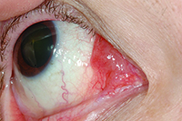
External examination revealed no evidence of proptosis and normal eyelids without edema or ptosis. Slit-lamp examination of the right eye revealed a painless mass in the medial canthus, involving the conjunctival stroma, appearing “salmon pink,” and measuring approximately 15 mm x 15 mm (See Figure 1). The left eye was unremarkable. Dilated fundus examination revealed normal optic nerve, macula, vessels and peripheral retina bilaterally.
Please click this link for diagnosis, workup, treatment and discussion.


