Mechanism of Retinal Detachment
The mechanism of rhegmatogenous retinal detachments centers on the relationship between the vitreous and the retina. The key event that can predispose an eye to retinal tears and detachment is a posterior vitreous detachment (PVD). With aging, the vitreous becomes syneretic (liquefied) and may detach itself from the retina. This detachment begins posteriorly and progresses anteriorly toward the vitreous base, and it is here that retinal flap tears (horseshoe tears) are commonly located. Retinal tears are found primarily in areas of strong vitreoretinal adhesion, such as the lateral and posterior border of lattice degeneration (See Figure 1). A retinal flap tear can progress to a retinal detachment because of increased movement of syneretic vitreous fluid though the open break and into the subretinal space. The continued vitreous traction on the anterior flap of the tear facilitates the process. A subclinical detachment is defined as a retinal break with surrounding subretinal fluid extending at least one disc diameter (DD) away from the break, but no more than two DD posterior to the equator. If there is less than one DD of fluid, then it is considered a retinal break without detachment.2
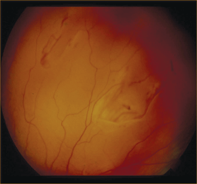 |
| Figure 1. Peripheral retinal breaks. |
Surgical Goals
In order to effectively treat a retinal detachment, all retinal breaks should be identified and treated to create chorioretinal adhesion, and causative vitreoretinal traction needs to be relieved. Creation of chorioretinal adhesion is performed with either laser photocoagulation or transconjunctival cryotherapy. Vitreoretinal traction is relieved either by removing the vitreous itself (by pars plana vitrectomy) or indenting the sclera so that the retinal break is reapproximated to the retina (by scleral buckle).
Pneumatic Retinopexy
Pneumatic retinopexy (PR) involves an intravitreal injection of gas (typically 0.3 cc of pure gas) combined with chorioretinal adhesion to the retinal break by either transconjunctival cryotherapy or laser photocoagulation. This is followed by a short course of appropriate post-procedure head positioning. The principle is that the buoyancy and surface tension of the intraocular gas bubble, which is typically sulfur hexafluoride (SF6) or perfluoropropane (C3F8), applies upward pressure to the retina and closes the retinal break. The chorioretinal adhesion promotes reattachment of the retina, with the retinal pigment epithelium pump responsible for removing the subretinal fluid. Since head positioning with intraocular gas can be tedious, PR has been advocated for retinal detachments caused by retinal breaks in the superior retina spanning less than one clock hour in size, thus allowing the patient to maintain a primarily face-down position, with some degree of head tilt, if necessary.
Contraindications for PR include uncontrolled glaucoma, presence of proliferative vitreoretinopathy, and a physical or mental disability which would not allow for appropriate postoperative head positioning. An advantage of this technique is the ability to perform this procedure in the office, which avoids the high cost and scheduling limitations of the operating room. Additionally, PR may be especially useful in cases where there are posterior breaks that are difficult to treat with scleral buckling and in cases where a filtering bleb is present or planned, since PR requires no significant manipulation of the conjunctiva. Also, patients who have PR performed have less postoperative pain and have quicker recovery times. Failure of PR does not preclude subsequent scleral buckling or vitrectomy surgery and it does not adversely impact visual outcome.3 However, visualization during the secondary surgery may be impaired due to residual gas and the detachment may be more complicated because of new breaks that can be induced by pneumatic retinopexy. Although not performed routinely because of difficulties with postoperative positioning, PR has been performed in cases involving inferior retinal breaks.4,5
One comprehensive review of the literature described 1,274 eyes treated with pneumatic retinopexy, and found a single-operation success rate of 80 percent.6 PR has typically been performed in phakic individuals but data from the Retinal Detachment Study Group suggests no difference in outcomes based on the status of the lens.7
Scleral Buckle
Scleral buckle surgery, in conjunction with chorioretinal adhesion, is an effective technique for the repair of rhegmatogenous retinal detachments. This technique works by altering the geometry and fluid dynamics of the eye such that closure of retinal tears is achieved. Most commonly, scleral buckles used today consist of solid silicone rubber, although other materials that are available include silicone sponges and fascia lata. The solid silicone rubber, commonly termed an explant, has a segmental component with an accompanying element that encircles the eye and is secured to the sclera with partial thickness scleral sutures or by passing the explant through partial-thickness scleral tunnels. The scleral buckle is placed so that once the retina is flattened, the breaks will lie upon the area of indentation created by the scleral buckle. Flattening of the retina can be achieved by creating the chorioretinal adhesion and then either relying on the retinal pigment epithelial pump to remove subretinal fluid or by manually performing external drainage of subretinal fluid. With this method, a full-thickness scleral dissection is performed and after micropuncture of the choroid, subretinal fluid egresses outside of the eye.
Scleral buckling is an external eye procedure and avoids the well-recognized complications of intraocular surgery. It causes minimal progression of cataract, which tends to be an important issue in younger patients. Also, as it is not always an intraocular procedure, the relative risk of endophthalmitis is decreased. However, scleral buckling involves a fair amount of conjunctival dissection and this may be of concern in patients with coexisting glaucoma. There is typically greater postoperative pain and there is a risk of infection or extrusion of the buckling element. Disadvantages also include induced myopia, diplopia, risk of inadvertent scleral perforation and complications related to external drainage of subretinal fluid. With external drainage, potential complications include choroidal hemorrhage and incarceration of the retina into the drainage site. To minimize these risks, segmental buckling combined with nondrainage procedures may be helpful.
Single operation anatomic success rate with SB has been reported to be 82 percent and overall success rate is 95 percent.8 A lower success rate is found in pseudophakic patients and in those cases where a retinal break is not found. This may be due to smaller peripheral breaks in pseudophakic eyes, which can often be often obscured by capsular opacities. A randomized clinical trial compared scleral buckling to pneumatic retinopexy and found a similar single-operation anatomic success rate of 73 percent and 82 percent, respectively.9
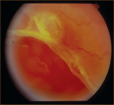 |
| Figure 2. Retinal detachment due to retinal dialysis. |
Pars Plana Vitrectomy
Standard 20-ga. pars plana vitrectomy (PPV) involves removing the vitreous and relieving primary vitreoretinal traction. The subretinal fluid is drained internally through the retinal break or through a separately created drainage retinotomy. After the retina is flattened, endolaser photocoagulation is used to create chorioretinal adhesion. An intraocular gas bubble or silicone oil can be used to provide tamponade to the retinal breaks in the postoperative period.
Many types of detachments are not amenable to repair by scleral buckling and need to be dealt with vitrectomy, such as those with giant retinal tears, proliferative vitreoretinopathy, posterior retinal breaks, and breaks with significant vitreous hemorrhage (See Figure 2). Advantages of PPV include direct removal of the vitreous and the ability to flatten the retina intraoperatively, without the risks of external subretinal fluid drainage. There is typically less postoperative pain with vitrectomy as compared to scleral buckling. Also, there is less induced myopia and manipulation of the conjunctiva with PPV. Vitreous floaters can persist with SB surgery, but these opacities can be removed in vitrectomy. By using intraoperative non-contact wide-angle viewing systems, small peripheral retinal breaks can be visualized easily and a thorough shaving of the vitreous base can be performed in pseudophakic patients.10 Also, in eyes with capsular fibrosis, vitrectomy can allow for improved visualization to the posterior segment. However, vitrectomy can cause progression of the cataract, so it tends to be preferred in pseudophakic patients. Also, placement of an intraocular gas bubble can provide significant short-term visual limitations, especially in monocular patients. PPV is associated with an increased risk of iatrogenic breaks, endophthalmitis, and postoperative PVR.11
Anatomic reattachment in primary vitrectomy for retinal detachments in phakic and pseudophakic patients ranges from 84 percent to 92 percent.12 Thus, in terms of visual success, primary vitrectomy seems to be as effective as scleral buckling for rhegmatogenous retinal detachment.
Combined PPV and Scleral Buckle
At times, combined PPV/SB may be indicated to repair a primary retinal detachment. In these situations, a solid silicone encircling element is placed around the eye to support the posterior vitreous base and a vitrectomy is performed and drainage of subretinal fluid is performed internally. The PPV/SB can be used in situations where there is widespread peripheral pathology associated with a retinal detachment. The encircling band provides support to the vitreous base, while vitrectomy removes the direct vitreoretinal traction that is present.
A surgical dilemma exists for repairing inferior retinal detachments. Primary vitrectomy with intraocular gas tamponade has been thought to be ineffective for inferior detachments since gas tamponade would be limited inferiorly. Combination PPV/SB may provide support to the inferior pathology. However, we have recently completed a study of patients treated with SB or combined PPV/SB for primary inferior rhegmatogenous retinal detachments and found no difference in their final visual outcome.13
Our Approach
Although there are various algorithms for the repair of primary retinal detachments, our approach is as follows. Pneumatic retinopexy is used for phakic individuals with superior detachments with isolated breaks. For larger primary detachments in a phakic individual with clearly visible retinal breaks, scleral buckling is our preferred treatment of choice. We perform external drainage of subretinal fluid in cases of largely bullous detachments. Primary vitrectomy is reserved for individuals with giant retinal tears, posterior pathology, or vitreous hemorrhage. We prefer vitrectomy in pseudophakic individuals, since the issue of cataract progression is absent and more importantly, a more thorough peripheral vitrectomy can be performed, without risk of damaging a crystalline lens. We prefer combined PPV/SB in certain cases of pseudophakic detachments with a large number of retinal breaks and an increased risk of proliferative vitreoretinopahy. We use either primary SB or combined PPV/SB in cases of inferior retinal detachments, where the encircling band can support the posterior vitreous base inferiorly, and in cases of widespread peripheral lattice degeneration.
Surgical Experience
The decision of the treatment method to use in repairing a retinal detachment is often affected by the surgeon's experience and comfort level with these techniques. Scleral buckling surgery has been considered the primary method of repair of retinal detachment. However, in recent years, pneumatic retinopexy and primary vitrectomies have become increasingly popular. Newer instrumentation in vitrectomy surgery has allowed for a more efficient surgical repair with increased comfort to the patient. With conventional scleral buckles being performed less frequently in their training, younger vitreoretinal surgeons tend to gain greater experience with PR and PPV and may have a bias when deciding on a treatment method for repairing a retinal detachment. A recent study demonstrated the popularity of pneumatic retinopexies was inversely proportional to the number of years a vitreoretinal surgeon has been in practice.14
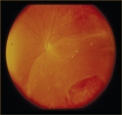 |
| Figure 3. An inferior retinal detachment. |
Timing of Intervention
One of the key findings to note in retinal detachments is if the macula is attached (macula-on) or macula-detached (macula-off). It is the status of the macula that defines the urgency with which surgical repair should be undertaken. Macula-on detachments are considered vision-threatening and in order to prevent extension of the detachment to involve the macula, they are considered surgical urgencies. Most vitreoretinal surgeons feel comfortable in delaying surgery a short time for uncomplicated macula-on retinal detachments, provided operating time is available the following day. If a delay is anticipated for logistical or medical reasons, strict bed rest and bilateral patching can delay progression of the retinal detachment into the macula. If the detachment is macula-off, surgery can be delayed for up to 10 days, without compromising final visual outcomes (See Figure 4).
Historically, all retinal detachments, including both macula-on and macula-off detachments, were considered surgical emergencies and were taken for immediate surgical repair, regardless of the time of day. The prevalent thinking at that time was that this type of repair could lead to improved outcomes instead of delaying the surgery. More recent studies have not demonstrated improved outcomes with immediate surgery on both types of retinal detachments.15-18 Surgery performed after-hours can involve working with operating room staff with limited retina experience. Emergency surgery is about 25-percent more costly than scheduled surgery, primarily because of higher wages of support staff in off hours.
New Frontiers
There is constant advancement in the technology and instrumentation that retinal surgeons use. This has led to improved visual outcomes and decreased operative times, with 25-ga. vitrectomy as an example. In this system, sclerotomies are placed through the intact conjunctiva with a trocar used for the infusion, light pipe, and surgical instrument. At the conclusion of the case, the trocars are removed and the wounds self-seal without the need for sutures. Advantages of this system include less postoperative astigmatism, since suturing of sclerotomies is typically not performed, and less inflammatory response of the eye. Also, there is less operating time since opening and closing of sclerotomies and conjunctiva are not required. Overall, this leads to a quicker visual recovery for the patient. However, since 25-ga. surgery is smaller, the instruments are less rigid than ones used in standard 20-ga. surgery, and moving the eye with the instruments can be challenging. This can become a limiting factor in removing peripheral vitreous and repairing complex retinal detachments. Also, the light source for 25-ga. instruments is less bright since there are less lumens available for transmission through a smaller-gauge fiber-optic light pipe. In addition, 25-ga. sclerotomies can leak at the end of surgery, requiring placement of sutures, and can be associated with postoperative hypotony.
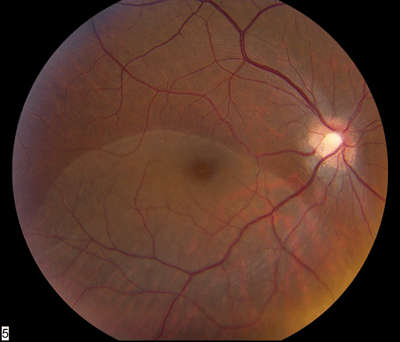 |
| Figure 4. A macula-off retinal detachment due to a peripheral break (not visible.) |
To alleviate issues of decreased light and rigidity, 23-ga. vitrectomy was developed. 23-ga. vitrectomy provides more rigid instrumentation with improved light transmission, thus allowing greater rotation of the eye and the ability to perform a more complete vitrectomy. The sclerotomies in this system are also done with a trocar transconjunctival system, but with beveled self-sealing incisions. Advances in retinal surgical instrumentation, by way of the 25-ga. and 23-ga. vitrectomy, add additional tools in the armamentarium of the vitreoretinal surgeon.
The choice and timing of the surgical technique to use in repairing a primary retinal detachment depends on the clinical scenario and the judgment of the surgeon. Unfortunately, even a randomized controlled clinical trial may not answer the question of which treatment method is the best, since each detachment is associated with its own dilemmas. However, thankfully vitreoretinal surgeons have been able to obtain a high success rate with all of these techniques and it is our hope that with improved technology and instrumentation, these outcomes can be improved further.
Dr. Tewari is a vitreoretinal fellow at the Barnes Retina Institute,
Washington University Department of Ophthalmology and Visual Science. Dr. Shah is from the Barnes Retina Institute and is a clinical associate professor with Washington University Department of Ophthalmology and Visual Science. Contact Dr. Shah at Barnes Retina Institute, 1600 S. Brentwood Blvd., Suite 800, St. Louis, Mo., 63144. Phone: (314) 367-1278, fax: (314) 962-2770, e-mail: gkshah1@gmail.com.
1. Wilkes SR, Beard CM, Kurland LT, et al. The incidence of retinal detachment in Rochester, Minn., 1970-1978. Am J Ophthalmol 1982;94:670-3.
2. Davis MD. Natural history of retinal breaks without detachment. Arch Ophthalmol 1974;92:183-94.
3. Ambler JS, Meyers SM, Zegarra H, Paranandi L. Reoperations and visual results after failed pneumatic retinopexy. Ophthalmology 1990;97:786-90.
4. Mansour AM. Pneumatic retinopexy for inferior retinal breaks. Ophthalmology 2005;112:1771-6.
5. Chang TS, Pelzek CD, Nguyen RL, et al. Inverted pneumatic retinopexy: a method of treating retinal detachments associated with inferior retinal breaks. Ophthalmology 2003;110:589-94.
6. Hilton GF, Das T, Majji AB, Jalali S. Pneumatic retinopexy: principles and practice. Indian J Ophthalmol 1996;44:131-43.
7. Tornambe PE, Hilton GF. Pneumatic retinopexy. A multicenter randomized controlled clinical trial comparing pneumatic retinopexy with scleral buckling. The Retinal Detachment Study Group. Ophthalmology 1989;96:772-83.
8. Schwartz SG, Kuhl DP, McPherson AR, et al. Twenty-year follow-up for scleral buckling. Arch Ophthalmol 2002;120:325-9.
9. Tornambe PE, Hilton GF. Pneumatic retinopexy. A multicenter randomized controlled clinical trial comparing pneumatic retinopexy with scleral buckling. The Retinal Detachment Study Group. Ophthalmology 1989;96:772-83.
10. Spitznas M. A binocular indirect ophthalmomicroscope (BIOM) for non-contact wide-angle vitreous surgery. Graefe's Arch Clin Exp Ophthalmol 1987;225:13-5.
11. Cowley M, Conway BP, Campochiaro PA, et al. Clinical risk factors for proliferative vitreoretinopathy. Arch Ophthalmol 1989;107:1147-51.
12. Campo RV, Sipperley JO, Sneed SR, et al. Pars plana vitrectomy without scleral buckle for pseudophakic retinal detachments. Ophthalmology 1999;106:1811-5.
13. Tewari A, Shah G.K., Patel A, et al. Management of Inferior Rhegmatogenous Retinal Detachments: Scleral Buckle vs. Combined Pars Plana Vitrectomy and Scleral Buckle. Submitted for publication . 2006.
14. Benson WE, Chan P, Sharma S, et al. Current popularity of pneumatic retinopexy. Retina 1999;19:238-41.
15. Yang CH, Lin HY, Huang JS, et al. Visual outcome in primary macula-off rhegmatogenous retinal detachment treated with scleral buckling. J Formos Med Assoc 2004;103:212-7.
16. Hassan TS, Sarrafizadeh R, Ruby AJ, et al. The effect of duration of macular detachment on results after the scleral buckle repair of primary, macula-off retinal detachments. Ophthalmology 2002;109:146-52.
17. Ross WH. Visual recovery after macula-off retinal detachment. Eye 2002;16:440-6.
18. Hartz AJ, Burton TC, Gottlieb MS, et al. Outcome and cost analysis of scheduled versus emergency scleral buckling surgery. Ophthalmology 1992;99:1358-63.


