Presentation
A 22-year-old man presented to the Wills Eye Hospital Emergency Room with worsening pain and swelling of his left upper eyelid that began three days prior to evaluation. The patient reported sinus congestion with yellow-green drainage for at least one month. He also noted poor appetite and fevers, and was unable to open his eye on the day of presentation. The patient initially presented to an outside emergency room where he was given intravenous antibiotics. A lateral canthotomy was attempted, and the patient was then transferred to the Wills Eye Hospital Emergency Room.
Medical History
The patient had a past medical history of chronic sinus infections. He otherwise denied any significant medical, surgical or ocular history. There was no pertinent family history, allergies or social history, and the patient was not taking any medications.
Examination
On initial examination, the patient was found to have a fever of 38.3 degrees Celsius (100.9 F), heart rate of 77 beats per minute, blood pressure of 158/74 mmHg, and a normal oxygen saturation on room air. Ocular examination revealed
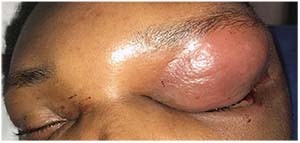 |
| Figure 1. External photography of the patient on initial presentation showing severe upper-lid edema and erythema. |
uncorrected visual acuity of 20/20 in the right eye and hand motion without improvement with pinhole in the left. Visualization of the left eye was severely limited by upper-lid edema, but pupils appeared round and reactive without obvious relative afferent pupillary defect and extraocular motility was grossly full. Intraocular pressure was 18 mmHg in the right eye, and greater than 70 mmHg in the left eye by Tono-Pen.
Anterior examination of the left eye showed tense upper-lid edema with tenderness and erythema, mild lower-lid edema and a cut lateral canthus (Figure 1). The globe appeared grossly formed with diffuse conjunctival injection and hemorrhagic chemosis greatest laterally and inferiorly. Desmarres retractors were used to evaluate the eye and further examination was otherwise normal. Anterior and posterior examination of the right eye was within normal limits.
Click here for Workup, Diagnosis & Discussion.


