"I was looking for something that was battery-powered and could be used for patients on stretchers, in beds or in the ER," he explains, "any setting where you can't get them to a standard fundus camera like the Topcon." He also envisions the camera making it easier for doctors' offices to screen patients for diabetic retinopathy, since he says a normal office isn't going to pay tens of thousands for a unit they use for only 20 patients a month.
 His most advanced prototype is a mydriatic, two-piece design consisting of a Panasonic Lumix G2 digital camera attached to a novel optical attachment he's devised that contains the illumination source and an image mask. It uses a xenon flash that he's detached from its normal place on the camera so it's in a better position to illuminate the retina. The system focuses automatically using the camera's built-in mechanism. The camera uses a front-mounted 22-D ophthalmic lens that gives a 60-degree field of view of the retina. It also has automatic through-the-lens metering, which sets the proper exposure for the camera in conjunction with the flash. Altogether, the non-camera parts cost less than $500. The camera cost is extra, but that's the idea. "That way, someone can buy a camera at Best Buy and snap it on the device," he says. He's quick to note, however, that better cameras, such as the Lumix (retail price between $650 and $700) will yield better shots. He and his co-engineer Ken Tran, a UVA undergrad, are currently looking for a partner to manufacture the system.
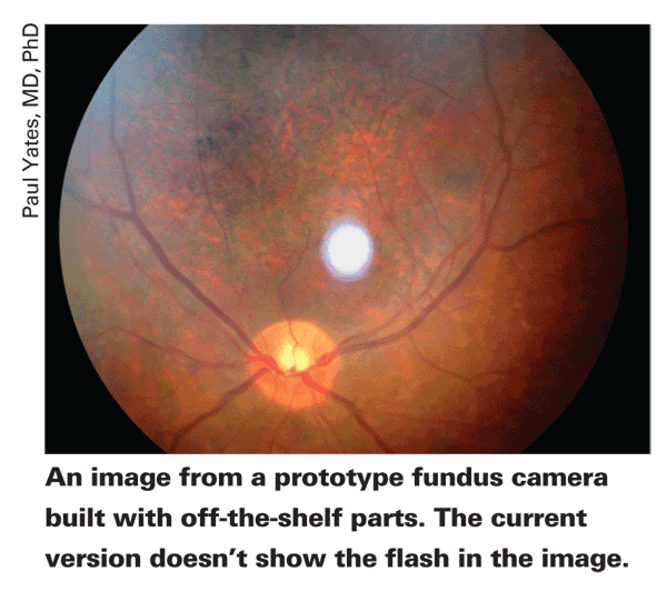
How are the images? "If you ask whether they look as good as a state-of-the art fundus camera, they don't," says Dr. Yates. "But they're as good as non-mydriatic cameras still in use today that came out in the 1990s. With screening, though, you're looking for dot and blot hemorrhages, exudates, et cetera. If you can see them, it's immaterial whether or not it's a super-high quality photo. The question is if any pathology is invisible or missing, and that's what we're studying in the clinic right now. However, the answer so far is that you pretty much get everything. There may be one or two cases where dot and blot hemorrhages may have been overlooked, but we think this will be a viable solution for screening purposes."


