The miniaturization of digital imaging technology that’s allowed cameras to be placed in cell phones and even smaller devices has made it possible to easily capture images wherever we go, and this revolution has now found its way to the ophthalmologist’s bag of tricks. If you have a patient who’s hard to move or is bedridden, or you’d just like to provide a patient an instant view of posterior segment pathology so he can understand it better, there are several new devices to choose from. Here’s a look at how they work.
|

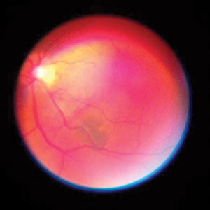
The EyeQuick offers a 10-degree field of view for retinal shots. The view is greater for external shots, because the farther you pull the camera away from the eye, the greater the field of view.
For a patient who has trouble keeping still for the ophthalmoscope image, the EyeQuick can capture a video. “You may want to capture the eye in video mode,” explains Dr. Ellman. “Then you’ll probably capture the image that you want from the video. For example, if you want a wider field of view, you can put it in video mode and sweep the area.” It has an adjustable white light, a green light for highlighting blood, a cobalt blue filter and a glare filter. “It’s optimized to take images under low light,” says Dr. Ellman. “So you actually capture them under a less intense light than you normally would with a direct ophthalmoscope. No flash is needed.”
The images are stored in JPEG format within the device (which has 220 MB of storage space). To transfer them elsewhere for e-mailing, printing or loading into an electronic health records program, there’s a USB port.
The EyeQuick, which retails for $5,995, has Food and Drug Administration 510(k) clearance as an imaging device, which means users can bill for pictures taken with it. Physicians use code 92250 for retinal photography (reimbursement for it is around $75) and code 92285 for anterior segment photography (reimbursement is around $25). When a unit is purchased, an EyeQuick representative will deliver it and provide training. There’s also a two-week evaluation period during which a physician can return the device for a full refund. For information, call 1 (800) 596-8335,
e-mail info@eyequick.com, or visit
eyequick.com or
facebook.com/eyequick.
• Keeler Portable Slit Lamp’s iPhone attachment. The PSL iPhone attachment allows the physician to remove one of the eyepieces of the PSL and replace it with a frame that holds an iPhone (sold separately). He then can use the iPhone’s still imaging and/or video applications to capture the view through the ocular. The PSL features 10X and 16X magnification, adjustable illumination, fixation targets and a 1-mm-square light patch for assessing anterior chamber flare. As a portable device, the user can put it in an optional $195 carrying case and take it out of the office for consults or hospital work. The PSL costs $3,995, and the iPhone adapter costs $195. For information, visit
keelerusa.com or call 1 (800) 523-5620.
• iExaminer/Welch Allyn PanOptic iPhone system. The iExaminer is an ophthalmic imaging application created for the iPhone 4 and iPhone 4S that’s designed to be attached to the PanOptic direct ophthalmoscope using a specially made mount.
The app and the mounting system were designed by Shreveport, La., ophthalmologist Wyche Coleman and his company Intuitive Medical Technologies. “The wide field of view is the reason we used the PanOptic for the device,” Dr. Coleman says. “You need to use a direct ophthalmoscope with it, and the PanOptic has a 26-degree field of view, even through an undilated pupil. The goal was to make a product that wasn’t just for ophthalmologists, but instead give all professionals access to the posterior segment.
|
“I go to a couple of satellite clinics and do mostly cataract evaluations,” Dr. Wyche continues. “At a clinic at which there’s no fundus camera, when I run across some pathology on a preop exam, such as a glaucoma suspect or someone with toxoplasmosis scars, it’s nice to be able to document it right there. And these are people you may never be able to get to your office and put in front of a fundus camera. Also, it’s helpful even in my main office where I have a fundus camera, in cases where I want to just show a patient what’s in the back of his eye and not have to get up and take him to the other room.”
The system’s resolution is based on the iPhone, so it can be 5 MP (iPhone 4) or 8 MP (iPhone 4S), and the iExaminer allows the user to toggle between a high- and low-res setting. It works by taking a series of images over a five-second period, to increase the odds of getting exactly the image you want. “If you use it in high-res to take a 5- or 8-MP shot, you get fewer shots to choose from,” explains Dr. Wyche. “If you’re looking at a child who can’t look straight for very long, you’re probably better off using the low-res setting, because it will take about 50 shots in that period of time. So, if at any point the patient is looking where you want him to look for 1/10 of a second, you’ll get something useful.”
After the series of shots is taken, the user scrolls through iExaminer and selects the image or images that are best for what he wanted to see; he can then save them with the patient’s demographic information. The app lets the user email the images along with some exam findings or print out the images using Apple’s AirPrint function. “For purposes of text messaging, you can save the iExaminer images to your iPhone camera roll and text message them from there,” says Dr. Coleman. “When you take a picture with a fundus camera in the office, it’s a process trying to export the image to do anything with it. It’s nice to be able to simply send a text message along with the image right from the iPhone after you capture it.”
The device’s battery life depends on the battery that’s used, adds Dr. Coleman. “We recommend using the new lithium-ion battery pack made by Welch Allyn,” he says. “I’ve never run one out, even after using it all day demonstrating the product on an exhibit hall floor.” He says you can also attach a conventional Welch Allyn battery handle, which fits in the charger in the ophthalmologist’s office.
Dr. Coleman is currently working toward 510(k) clearance for the device and expects the system to be available for sale in August 2012. For information, visit
iexam.com. REVIEW


