Presentation
A 76-year-old Caucasian male was referred to the Wills Eye emergency room for evaluation of blurred vision and diplopia after a fall. He had reportedly tripped over a hose, fell, and hit the left side of his face and his left eye on a lawn mower several hours prior to presentation. The patient was wearing safety glasses and didn’t lose consciousness during the fall. He initially presented to an outside emergency room where a left brow laceration was repaired, and CT scans of the brain and cervical spine were negative. He was referred for further evaluation of left orbital floor and medial wall fractures on maxillofacial CT scan.
Medical History
Two years prior to presentation, he was struck in the right eye by a high-tension wire while riding a tractor and suffered a vitreous hemorrhage and a retinal tear for which he underwent pars plana vitrectomy and laser retinopexy. Past ocular history also included cataract surgery and ocular hypertension in both eyes, and YAG capsulotomy in the right eye. Past medical history included hypertension, hyperlipidemia, type II diabetes mellitus, obstructive sleep apnea, Alzheimer’s disease, restless leg syndrome, osteoarthritis and benign prostatic hypertrophy. Social and family histories were non-contributory. Current medications included dorzolamide-timolol in both eyes, amlodipine, lisinopril, atorvastatin, aspirin, pioglitazone, niacin, quetiapine, pramipexole, memantine, galantamine, sertraline, tamsulosin and meloxicam.
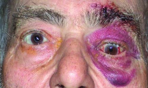 |
| Figure 1. External photo showing left periorbital ecchymosis and edema with hypoglobus in primary gaze after a |
Examination
Ophthalmic examination demonstrated a visual acuity of 20/40 OD (pinhole 20/20) and 20/25 OS (PH no improvement). Pupils and confrontation visual fields were normal, and Ishihara color plates were 8/8 OU. Motility of the right eye was full, while motility of the left eye demonstrated 0-percent supraduction, 95-percent adduction, 90-percent infraduction and 100-percent abduction. A right hypertropia was present in primary gaze (25 prism diopters), which worsened in upgaze (>30 prism diopters). Intraocular pressure by Goldmann tonometry was 14 mmHg in both eyes. External examination revealed sutured lacerations of the central forehead and left eyebrow, 360 degrees of ecchymosis and edema around the left orbit, and enophthalmos and hypoglobus of the left eye in primary gaze (Figure 1). There was no bony step-off palpated on external examination of the orbital rim. Hertel exophthalmometry demonstrated enophthalmos of the left eye with a base of 121 mm, 20.5 mm OD and 18 mm OS. Sensation was intact in the V1 and V2 distributions.
Slit lamp examination was notable for 1+ conjunctival injection, temporal subconjunctival hemorrhage, superior superficial punctate keratopathy of the left eye and posterior chamber intraocular lenses in both eyes. Dilated fundus examination was normal in both eyes. Forced duction and force generation testing were performed after instillation of viscous lidocaine. Forced duction testing was normal in the right eye and revealed restriction of vertical motility in the left eye. Force generation testing was normal in both eyes.
What is your diagnosis? What further workup would you pursue?
Please click this link for diagnosis, workup, treatment and discussion.


